
Introduction: When Knee Pain Takes Over Your Life
You wake up in the morning. Before your feet even touch the floor, it starts – that deep, grinding ache in your knee. Stairs feel like a test of willpower. A simple walk to the kitchen becomes something you dread. Over time, you stop doing the things that matter most: morning walks, playing with your grandchildren, standing long enough to cook a full meal, or even sitting through a movie without shifting uncomfortably.
If this is your life right now, you are not alone.
Chronic knee pain caused by arthritis, joint degeneration, or old injuries affects millions of people across India – and across Mumbai in particular. For decades, the only lasting solution was traditional total knee replacement, a major surgery with a long recovery that many patients feared.
That has changed.
Minimally Invasive Total Knee Replacement (MI-TKR) has emerged as one of the most significant advances in modern orthopedic surgery. It offers the same permanent relief from knee pain as traditional surgery – but with a smaller incision, less damage to surrounding tissue, less post-operative pain, and a faster road back to your normal life.
At our practice, Dr. Abhay Chhallani performs this advanced procedure for patients from across Mumbai, Navi Mumbai, and Maharashtra. As a leading specialist in Robotic Knee Replacement in Navi Mumbai, Dr. Chhallani helps patients get back to walking, moving, and living – often faster than they ever expected.
This comprehensive guide will walk you through everything: what the surgery involves, how it is performed, who is a good candidate, what the different types of implants are, how to prepare before surgery, what recovery looks like week by week, the risks to know about, and how to choose the right surgeon.
What Is Minimally Invasive Total Knee Replacement?
Minimally Invasive Total Knee Replacement – also written as MI-TKR or minimally invasive total knee arthroplasty (MI-TKA) – is an advanced form of the standard total knee replacement procedure.
The fundamental goal is identical to traditional surgery: the damaged surfaces of the knee joint are removed and replaced with precision-engineered metal and high-density plastic implants. This eliminates the bone-on-bone grinding that causes chronic knee arthritis pain and restores smooth, stable joint movement.
What is different is the surgical approach.
In traditional knee replacement surgery, the surgeon makes a large incision of 8 to 12 inches down the front of the knee and fully cuts through the quadriceps tendon – the powerful tendon that connects the thigh muscles to the kneecap. The kneecap is then flipped 180 degrees and the shinbone (tibia) is often dislocated to fully expose the joint.
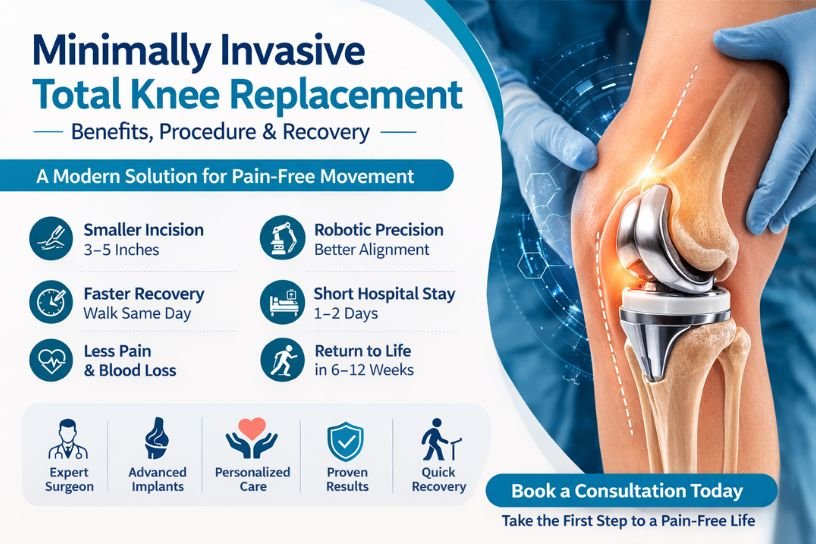
In minimally invasive knee replacement, the incision is just 3 to 5 inches. Rather than cutting through the quadriceps tendon, the surgeon gently moves the muscles and soft tissue aside. The kneecap is shifted rather than flipped. The tibia is usually left in place.
This approach is known as “quadriceps-sparing” – and it is the key reason why recovery is faster and post-operative pain is lower.
The implants used are identical. Only the technique changes.
Understanding the Knee Joint: Why It Breaks Down
To understand why knee replacement becomes necessary, it helps to understand how the knee works and why it can fail over time.
The knee is the largest and one of the most complex joints in the human body. It connects three bones – the femur (thigh bone), the tibia (shin bone), and the patella (kneecap). The ends of these bones are covered with a smooth, white tissue called articular cartilage, which allows them to glide against each other without friction.
Between the femur and tibia sit two C-shaped pieces of cartilage called the menisci, which act as shock absorbers and help distribute weight evenly across the joint.
When articular cartilage breaks down – due to age, wear and tear, injury, or inflammatory conditions – the bones begin rubbing directly against each other. This causes:
- Constant or intermittent pain, especially with movement
- Swelling and stiffness in the joint
- A grinding, clicking, or locking sensation
- Progressive loss of mobility and flexibility
- Visible deformity such as the leg bowing inward or outward
Once cartilage is lost, it does not grow back on its own. This is why knee arthritis is progressive – it gets worse over time without treatment, not better.
Types of Knee Conditions That Lead to Replacement Surgery
Not all knee pain leads to replacement surgery, but several specific conditions are its most common causes:
Osteoarthritis (OA)
The most common cause of knee replacement surgery in India. Osteoarthritis is a degenerative joint disease where cartilage gradually wears away over time. It is more common with age, and women are more frequently affected than men due to differences in joint structure and hormonal factors. OA causes progressive pain, stiffness, swelling, and eventually bone-on-bone contact that makes even resting painful.
Rheumatoid Arthritis (RA)
An autoimmune condition in which the body’s immune system attacks the lining of the joint (synovium), causing inflammation, pain, and joint destruction. Rheumatoid arthritis can affect people of any age and often affects multiple joints at once.
Post-Traumatic Arthritis
Develops after a significant knee injury – such as a fracture of the knee bones, a torn ACL (anterior cruciate ligament), or a meniscal tear. Even injuries that appeared to heal well can lead to arthritis years or decades later as the cartilage degrades due to the mechanical disruption caused by the original trauma.
Avascular Necrosis (AVN)
A condition where the blood supply to the bone is cut off, causing bone tissue to die. In the knee, this typically affects the femoral condyle. Without adequate blood supply, the bone weakens and collapses, destroying the joint surface. AVN can be caused by long-term steroid use, excessive alcohol consumption, or prior trauma.
Severe Bone Deformity
Some patients develop significant bowing of the legs – either inward (varus deformity) or outward (valgus deformity) – due to uneven cartilage loss and bone wear. This deformity places abnormal stress on the knee and accelerates joint damage.
Who Needs Total Knee Replacement Surgery?
Total knee replacement surgery is not the first line of treatment – it is recommended only when conservative, non-surgical approaches are no longer providing meaningful relief.
You may be a candidate for total knee replacement surgery if:
- You have been diagnosed with severe knee osteoarthritis, rheumatoid arthritis, or post-traumatic arthritis
- You experience persistent knee pain that limits your everyday activities – walking, climbing stairs, rising from a chair, or sleeping
- Your knee is stiff for more than 30 minutes after waking up or after prolonged sitting
- X-rays confirm significant joint space narrowing, bone spurs, or bone-on-bone contact
- Your knee has noticeable deformity – bowing in or out
- Non-surgical treatments such as pain medication, physiotherapy, steroid injections, PRP therapy, and bracing have failed to provide adequate relief
- Your quality of life is seriously compromised by knee pain
- You are aged between 55 and 80 (though younger and older patients are also treated when clinically indicated)
The decision to proceed with surgery is never made lightly. It involves a thorough clinical evaluation, detailed imaging, a review of your overall health, and an open conversation between you and your orthopedic surgeon about your goals and expectations.
Non-Surgical Knee Arthritis Treatment Options
Before considering surgery, a well-structured non-surgical treatment plan is always the first step. Understanding these options helps patients know when they have genuinely exhausted conservative management.
Pain and Anti-Inflammatory Medications NSAIDs (non-steroidal anti-inflammatory drugs) such as ibuprofen or diclofenac reduce inflammation and manage pain. These are effective for mild to moderate arthritis but become less reliable as the condition progresses. Long-term NSAID use also carries risks for the stomach, kidneys, and cardiovascular system.
Physiotherapy and Exercise Therapy A structured physiotherapy program strengthens the muscles around the knee – particularly the quadriceps, hamstrings, and calf muscles – which reduces stress on the joint. Low-impact activities like swimming, cycling, and water aerobics are particularly beneficial for knee arthritis patients.
Weight Management Excess body weight dramatically increases the load on the knee joint. Studies show that each kilogram of body weight reduction reduces approximately 4 kilograms of force on the knee. Even modest weight loss can meaningfully reduce pain and slow the progression of arthritis.
Corticosteroid Injections Injections of corticosteroid medication directly into the knee joint can provide significant but temporary pain relief – typically lasting weeks to a few months. Most surgeons limit these to 3 to 4 injections per year to prevent further joint damage from the steroid itself.
Hyaluronic Acid (Viscosupplementation) Injections Hyaluronic acid is a naturally occurring substance in joint fluid that acts as a lubricant and shock absorber. Injectable hyaluronic acid supplements this fluid, potentially reducing pain and improving mobility. Results vary between patients.
Platelet-Rich Plasma (PRP) Therapy PRP is derived from the patient’s own blood. After centrifugation, the platelet-rich fraction is injected into the knee to stimulate tissue healing and reduce inflammation. PRP is increasingly used for early to moderate arthritis and can delay the need for surgery in appropriate patients.
Bracing and Orthotics Knee braces designed for arthritis patients can offload pressure from the damaged compartment of the knee, reducing pain and improving walking comfort. Custom shoe insoles (orthotics) can also correct biomechanical imbalances that worsen knee pain.
When these options no longer provide adequate relief, and when knee pain is significantly impacting your daily function and quality of life, total knee replacement surgery becomes the most effective long-term solution.
Minimally Invasive vs. Traditional Knee Replacement: A Detailed Comparison
Understanding the differences between the two approaches helps patients make informed decisions.
| Feature | Traditional TKR | Minimally Invasive TKR |
| Incision Length | 8–12 inches | 3–5 inches |
| Quadriceps Tendon | Cut through | Preserved / gently moved |
| Kneecap (Patella) | Flipped 180 degrees | Gently shifted to the side |
| Tibia Dislocation | Often required | Usually not required |
| Tissue Disruption | Significant | Minimal |
| Blood Loss | More | Less |
| Hospital Stay | 3–5 days | 1–2 days (or same-day) |
| Early Recovery | Slower | Faster in first 4–6 weeks |
| Long-Term Outcomes | Excellent | Equally excellent |
| Implants Used | Proven standard | Same proven standard |
| Surgical Difficulty | Standard | More technically demanding |
| Suitable For | Most patients | Carefully selected patients |
Important note: The long-term outcomes and implant durability of minimally invasive knee replacement are equivalent to traditional surgery. The advantages are primarily in the early recovery period – less pain, faster return of mobility, and shorter hospital stay.
Types of Minimally Invasive Knee Replacement Surgical Approaches
Orthopedic surgeons use several distinct techniques within the category of minimally invasive knee replacement. Each has slightly different incision placement and muscle handling:
1. Quadriceps-Sparing (QS) Approach
The most conservative minimally invasive technique. The incision avoids the quadriceps tendon entirely. The muscles are retracted – not cut – to access the joint. This approach offers the greatest quadriceps preservation but also provides the most limited surgical field, requiring exceptional skill and specialized instruments.
2. Mini-Midvastus Approach
A small incision is made in the vastus medialis oblique (VMO) muscle – one of the quadriceps muscles on the inner thigh. This gives slightly better access than the quadriceps-sparing technique while still avoiding the main quadriceps tendon. It is the most commonly used minimally invasive approach.
3. Mini-Subvastus Approach
The incision is placed below the vastus medialis muscle rather than through it, going underneath the muscle to reach the joint. This approach fully spares the quadriceps mechanism and can result in excellent early recovery of quad strength, but it is technically more challenging in patients with limited surgical access.
4. Jiffy Knee Technique
A newer, highly specialized approach where only the skin and joint capsule are incised. Specialized instruments gently lift and move the muscles aside without any muscle cutting at all. This technique is performed by very few highly specialized surgeons and is at the frontier of minimally invasive knee surgery.
Your surgeon will determine which approach is most suitable based on your anatomy, body type, and the specific pattern of your knee arthritis.
Types of Knee Replacement Implants
The choice of implant is a critical decision that your surgeon makes based on your age, bone quality, activity level, and the specific anatomy of your knee. Understanding the main types helps patients have more informed conversations with their doctor.
By Fixation Method
Cemented Fixation The most commonly used method worldwide. A fast-curing acrylic bone cement (polymethylmethacrylate) is used to bond the metal implant components to the prepared bone surfaces. Cemented fixation provides immediate, reliable stability and is the gold standard for older patients, those with osteoporosis, or anyone with weaker bone quality.
Cementless Fixation The implant surfaces are coated with a porous or textured material that encourages the patient’s own bone to grow directly into and around the implant over time. This biological bonding can provide outstanding long-term durability. Cementless implants are generally preferred for younger, more active patients with good bone quality. The bone ingrowth process takes several weeks to months to complete. Recent research and large registry studies show that cementless knee replacement achieves survivorship rates exceeding 98% at 5 years.
Hybrid Fixation A combination approach where the tibial (shin bone) component is cemented and the femoral (thigh bone) component is cementless, or vice versa. Used when bone quality varies between the two surfaces.
By Bearing Design
Fixed-Bearing Implants The polyethylene (plastic) spacer is fixed firmly to the metal tibial tray and does not move independently. These are durable, reliable, and well-suited for less active patients or those with simpler knee mechanics.
Mobile-Bearing Implants (Rotating Platform) The polyethylene spacer can rotate a small amount within the metal tibial tray, mimicking the natural rotational movement of the knee. This design can improve range of motion slightly and reduce wear on the plastic component. Mobile-bearing implants are often preferred for younger, more active patients who require more natural knee kinematics.
By Constraint Level
Cruciate-Retaining (CR) Design The posterior cruciate ligament (PCL) is preserved during surgery. This design aims to maintain a more natural feel in the knee and can improve proprioception (the sense of joint position). Suitable when the PCL is intact and functioning.
Posterior-Stabilized (PS) Design A post-and-cam mechanism built into the implant replaces the function of the PCL, which is removed during surgery. PS designs provide more reliable stability and are preferred when the PCL is damaged, incompetent, or removed.
Your surgeon will choose the implant design that best matches your anatomy, lifestyle, and long-term goals.
Robotic Knee Replacement Surgery: The Future of Precision
One of the most exciting developments in knee replacement surgery is the integration of robotic-assisted technology with the minimally invasive approach.
Robotic knee replacement surgery – also known as robotic-assisted total knee arthroplasty – combines the soft-tissue-sparing benefits of minimally invasive surgery with extraordinary implant placement accuracy that goes beyond what can be consistently achieved with manual instrumentation alone.
Patients seeking Robotic Knee Replacement in Navi Mumbai now have access to this world-class technology close to home, with Dr. Abhay Chhallani offering robotic-assisted procedures using the latest surgical platforms available in the region.
How Robotic Knee Replacement Works
Before surgery, a detailed CT scan of the patient’s knee is used to create a precise three-dimensional model of the joint. Using this model, the surgeon and the robotic system together develop a fully customized surgical plan – specifying exactly where bone cuts will be made, what size implant will be used, and the ideal alignment and positioning of each component.
In the operating room, the robotic arm provides haptic feedback and real-time guidance to the surgeon. The arm can physically limit bone cuts to the pre-planned boundaries, making it virtually impossible to remove more bone than intended. This level of precision is particularly valuable in minimally invasive surgery, where the reduced field of vision makes accuracy more challenging.
Why Accuracy Matters So Much in Knee Replacement
The long-term success of a knee replacement depends heavily on how well the implant is aligned. Even a few degrees of misalignment can accelerate wear on the plastic spacer, reduce range of motion, cause ongoing pain, and ultimately shorten the life of the implant – potentially requiring revision surgery.
Robotic assistance helps ensure:
- Optimal implant alignment and positioning
- Correct leg axis and balance
- Precise soft tissue balancing
- Personalized implant sizing based on the patient’s exact anatomy
Computer-Assisted Navigation
For practices without robotic arms, computer-assisted navigation systems provide real-time 3D feedback during surgery using infrared sensors attached to the bones. Systems like OrthoPilot and Knee Track Module allow surgeons to verify alignment and implant position continuously during the procedure, significantly improving accuracy over conventional manual techniques.
Patient-Specific Instrumentation (PSI) Another technology where custom-designed surgical guides are manufactured from pre-operative CT or MRI scans. These guides fit the patient’s unique bone anatomy like a key in a lock, directing the exact location and angle of each bone cut. PSI reduces operating time and enhances reproducibility.
How to Prepare for Minimally Invasive Knee Replacement Surgery
The weeks and months before your surgery are just as important as the surgery itself. Patients who are well-prepared physically and mentally tend to recover faster and experience fewer complications.
4 to 8 Weeks Before Surgery: Pre-Habilitation Exercises
Pre-habilitation – strengthening the muscles around your knee before surgery – is one of the most evidence-backed ways to improve recovery outcomes.
Recommended exercises before knee replacement:
Quad Sets Lie flat on your back with your leg straight. Tighten the thigh muscle (quadriceps) and press the back of your knee into the floor or bed. Hold for 5 seconds, then relax. Repeat 10 times, twice daily.
Straight Leg Raises Lie on your back. Bend your non-operated leg with foot flat. Tighten the thigh of the operated leg and raise it to the height of the opposite knee. Hold for 3 seconds, lower slowly. Repeat 10 times.
Heel Slides Lie on your back. Slowly slide your heel toward your buttocks, bending the knee as far as comfortable. Hold for 5 seconds, then slide back. Repeat 10 times. This improves knee flexion range pre-operatively.
Short Arc Quads Place a rolled towel under your knee. Straighten your leg fully, hold 5 seconds, lower slowly. Repeat 10 times. Excellent for isolated quad strengthening.
Seated Knee Flexion and Extension Sit in a chair and bend and straighten your knee through its comfortable range of motion. Repeat 10–15 times. Especially beneficial before surgery to maintain range.
Standing Hip Abduction Stand holding a stable surface. Lift your leg out to the side, keeping it straight and toes forward. Hold 3 seconds, lower slowly. This strengthens the hip stabilizers, which support the knee.
Low-Impact Cardiovascular Exercise Stationary cycling, swimming, and water walking are excellent options before knee replacement. They build cardiovascular fitness, reduce pain, manage weight, and maintain muscle tone – all without placing excessive stress on the damaged joint.
Nutrition Before Knee Replacement Surgery
What you eat in the weeks before surgery directly affects your healing, immunity, and recovery speed.
High Protein Diet Aim to eat a high-quality protein source 3 to 4 times per day in the month before surgery. Protein is the building block of muscle and tissue repair. Good sources include chicken, fish, eggs, paneer, dal, lentils, and Greek yogurt.
Iron-Rich Foods Spinach, lentils, red meat, and fortified cereals support healthy red blood cell production, which is important both during surgery and healing.
Vitamin C Supports collagen production and wound healing. Found in citrus fruits, guava, amla, bell peppers, and tomatoes.
Calcium and Vitamin D Support bone health and strength. Dairy products, fortified foods, leafy greens, and safe sun exposure support calcium and vitamin D levels.
Fiber Constipation is a common side effect of post-operative pain medications (opioids). A fiber-rich diet before and after surgery – from whole grains, fruits, and vegetables – helps prevent this issue.
Hydration Stay well-hydrated in the days before surgery. Avoid alcohol for at least two weeks pre-operatively, as it can interfere with anesthesia, increase bleeding, and impair healing.
Other Pre-Surgery Preparation Steps
Stop Smoking Smoking significantly impairs healing by reducing blood flow and oxygen delivery to tissues. It also increases the risk of infection and wound complications. Stopping smoking at least 6 to 8 weeks before surgery substantially reduces these risks.
Medication Review Inform your surgeon about every medication and supplement you take – including blood thinners (aspirin, warfarin, clopidogrel), NSAIDs, diabetes medications, herbal supplements, and vitamins. Many of these need to be paused or adjusted before surgery.
Medical Clearance A pre-operative assessment with your physician will evaluate your heart health, lung function, blood sugar control (especially important for diabetic patients), and blood pressure. Any uncontrolled medical condition needs to be optimized before surgery can safely proceed.
Pre-Operative Tests Blood tests, electrocardiogram (ECG), chest X-ray, and knee imaging (X-rays and sometimes MRI) are typically required before surgery.
Home Preparation Arrange your home before surgery to make your recovery smoother:
- Remove loose rugs and tripping hazards from floors
- Set up a ground-floor sleeping area if stairs are involved
- Install grab bars in the bathroom
- Arrange a raised toilet seat if possible
- Stock up on easy-to-prepare meals for the first few weeks
- Arrange for someone to help you at home for the first 1 to 2 weeks
- If you have children or pets, arrange appropriate care
How Is Minimally Invasive Knee Replacement Surgery Performed? (Step-by-Step)
Pre-Operative Preparation
On the day of surgery, you will fast for 6 to 8 hours beforehand. An intravenous (IV) line will be placed, and antibiotic medication is given before the incision to prevent infection.
Anesthesia
The procedure is performed under general anesthesia (you are fully unconscious) or spinal anesthesia (you are awake but numb from the waist down). Many patients also receive a nerve block – an injection of local anesthetic around the knee nerves – which significantly reduces post-operative pain for the first 12 to 24 hours and reduces the need for strong opioid pain medications.
Tourniquet Application
A tourniquet is inflated around the upper thigh to temporarily reduce blood flow to the knee during the procedure. This reduces intraoperative blood loss and provides a clearer surgical field.
The Minimally Invasive Incision
A precise incision of 3 to 5 inches is made over the front of the knee. Using specially designed narrow retractors, the surgeon carefully moves the quadriceps muscle and soft tissue aside – rather than cutting through them.
Exposing the Joint
The kneecap (patella) is gently moved to one side rather than flipped over, limiting the disruption to the extensor mechanism of the knee. Specialized instruments allow the surgeon to work within the smaller field of view.
Bone Preparation
The surgeon precisely removes the damaged cartilage and a thin layer of underlying bone from the end of the femur, the top of the tibia, and if required, the undersurface of the patella. Specially designed minimally invasive cutting guides – or robotic/computer assistance – ensure these bone cuts are made at exactly the right angles.
Trial Components
Before cementing the permanent implants, trial components are placed to test the fit, alignment, range of motion, and ligament balance. This is one of the most critical steps – ensuring the knee feels natural and balanced before the final implants are secured.
Implant Fixation
The permanent metal femoral and tibial components are fixed using bone cement, cementless press-fitting, or a combination of both – depending on the patient and the implant selected. A high-quality polyethylene spacer is snapped between the two metal components. If the patella is being resurfaced, a plastic button is cemented to its undersurface.
Irrigation and Closure
The joint is thoroughly washed out. A drain may be placed to collect fluid post-operatively. The layers of tissue are carefully closed in sequence, and the skin is closed with sutures or staples.
Total Procedure Time
The entire surgery typically takes 1.5 to 2.5 hours, depending on complexity.
Key Benefits of Minimally Invasive Knee Replacement Surgery
1. Smaller Incision and Less Visible Scar
At 3 to 5 inches, the incision is less than half the length of traditional surgery. Patients – particularly those conscious about surgical scars – appreciate this significantly.
2. Less Post-Operative Pain
The quadriceps-sparing approach means less tissue is cut and less healing is required of the surrounding muscles. Much of the intense pain after traditional knee replacement comes from the healing of the divided quadriceps tendon – which is largely avoided here. This translates to lower opioid requirements post-surgery, which itself reduces side effects like nausea, constipation, and drowsiness.
3. Faster Early Recovery
Because the quadriceps are preserved, muscle function returns faster. Patients are typically walking with a walker on the same day as surgery and often progress to a cane within 2 to 3 weeks – faster than with traditional surgery.
4. Shorter Hospital Stay
Most patients undergoing minimally invasive knee replacement are discharged after 1 to 2 days. Carefully selected patients can even be discharged the same day as surgery. This reduces hospital costs and the risk of hospital-acquired infections.
5. Less Blood Loss
The smaller incision and reduced tissue handling naturally result in less bleeding during surgery, reducing the likelihood of needing a blood transfusion.
6. Lower Infection Risk
Smaller incisions mean less wound surface area exposed during surgery and less dead tissue to become infected. This is especially meaningful for patients with diabetes, obesity, or conditions that slow healing.
7. Faster Return of Knee Range of Motion
Patients with preserved quadriceps muscles tend to achieve better early range of motion in physiotherapy, which is directly linked to better long-term outcomes.
8. Earlier Return to Daily Life
Patients typically return to light daily activities in 2 to 4 weeks, driving in 4 to 6 weeks, and office work in 3 to 6 weeks.
9. Equivalent Long-Term Outcomes
The long-term durability and success rates of minimally invasive knee replacement are equivalent to traditional knee replacement. Modern knee implants have survivorship rates exceeding 90 to 95% at 10 years – meaning most patients get a decade or more of excellent, pain-free function before any further intervention is needed.
Understanding the Risks and Complications
Any surgical procedure carries risks, and it is important that patients have an honest understanding of what these are. The overall rate of serious complications following total knee replacement – minimally invasive or traditional – is low, typically less than 5 percent when performed by an experienced, high-volume surgeon.
Possible complications include:
Blood Clots (Deep Vein Thrombosis / DVT) One of the most common post-operative concerns. Blood clots can form in the deep veins of the leg after surgery. To prevent this, patients are started on blood-thinning medications and encouraged to mobilize early. Signs include increased leg swelling, redness, and warmth.
Pulmonary Embolism (PE) A blood clot that travels to the lungs. This is a serious complication but is rare when proper DVT prevention measures are taken.
Infection Infection around the knee implant is a rare but serious complication. Surgeons administer antibiotics before, during, and after surgery to minimize this risk. Patients are advised to inform dentists and other doctors about their knee replacement before any invasive procedures in future, as bacteria from elsewhere in the body can travel to the implant.
Implant Loosening or Misalignment Over years or decades, implants can loosen from the bone or shift slightly out of alignment. Modern implant design, robotic assistance, and careful patient selection have reduced this risk significantly.
Nerve and Blood Vessel Injury The nerves and blood vessels around the knee are at small risk of injury during surgery. Experienced surgeons using specialized minimally invasive instruments and modern technology minimize this risk.
Stiffness (Arthrofibrosis) Some patients develop excessive scar tissue inside the knee joint after surgery, leading to stiffness and limited range of motion. Consistent physiotherapy is the best preventive measure.
Implant-Specific Complications
- Cementless implants may have a period of slight pain until bone ingrowth is complete
- Mobile-bearing implants carry a very small risk of polyethylene dislocation
Anesthesia Risks Like all surgeries requiring anesthesia, there are small risks related to cardiac events, stroke, and reactions to anesthesia agents. Pre-operative clearance and a thorough anesthesia evaluation minimize these risks.
Overall: The risks of knee replacement surgery must always be weighed against the significant quality-of-life benefits for patients who have exhausted all conservative treatment options. The vast majority of patients experience significant, lasting improvement in pain and mobility.
Minimally Invasive Knee Replacement: Who Is – and Is Not – a Candidate?
Good Candidates
- Generally healthy patients aside from their knee condition
- Moderate to severe knee arthritis without extreme bone loss
- No prior major open knee surgery on the same knee
- BMI under approximately 35 (excess weight makes the limited access more technically difficult)
- No severe knee deformity (moderate deformity can often still be addressed)
- Intact or only mildly compromised ligaments
Patients Who May Need Traditional Surgery Instead
- Significant obesity (high BMI) that limits surgical access
- Severe bone deformity requiring more extensive correction
- Previous major open knee surgery with significant scar tissue
- Very severe bone loss requiring structural bone grafting
- Complex revision (redo) knee replacement cases
- Medical conditions that significantly increase surgical risk
The decision between minimally invasive and traditional surgery is made individually for every patient. What matters most is not the size of the incision – but that the surgery is performed correctly, with well-positioned implants, by a surgeon with the training and experience to deliver consistent results.
Week-by-Week Recovery Guide After Minimally Invasive Knee Replacement
Day of Surgery (Day 0)
Within hours of waking from anesthesia, a physiotherapist will visit you. Under guidance, you will sit on the edge of the bed, and most patients take their first steps with a walker on the same day. The operated leg is elevated to reduce swelling. Ice packs are applied regularly. IV pain medication transitions to oral medications.
Days 1–3: Early Recovery in Hospital
The focus shifts to controlled walking, going up and down steps (a required skill before discharge), independent transfer in and out of bed, beginning basic knee exercises (quad sets, heel slides, ankle pumps), and wound care.
Ankle pumps – repeatedly flexing and pointing the foot – are done every hour while resting. This activates the calf muscles and helps prevent blood clots.
Week 1–2: Home Recovery Begins
You will likely use a walker or forearm crutches. The priority is managing swelling (elevation and ice packs), wound hygiene, and performing your prescribed home exercise program 2 to 3 times per day.
Most patients experience their worst swelling during this period – this is completely normal. Swelling can persist for weeks to months as the body heals.
A physiotherapist visits at home or you attend outpatient sessions – this varies based on your recovery plan.
Week 3–6: Building Range of Motion and Strength
By week 3, most patients can walk with a cane or independently for short distances. Physiotherapy intensifies – targeting knee flexion beyond 90 degrees, full knee extension, stair climbing technique, and progressive muscle strengthening.
Driving is generally not recommended during this period, especially if the operated leg is needed for braking.
Most patients can resume light cooking, personal hygiene without assistance, and short outings.
Week 6–12: Growing Independence
The 6-week milestone is significant. Most patients have noticeably reduced pain, better walking endurance, and improved confidence in their knee. Low-impact activities like swimming and stationary cycling are typically introduced at this stage.
Office-based workers often return to work in this period. Driving is usually permitted after clearance from your surgeon, typically around 6 weeks.
3 to 6 Months: Return to Normal Life
By 3 months, the majority of patients feel dramatically better than before surgery. Pain is minimal or absent during most activities. Physiotherapy continues but decreases in frequency.
Activities like golf, light hiking, dancing, cycling, and recreational walking are generally achievable by this point for most patients.
6 Months to 1 Year: Full Recovery
Complete recovery – including the final return of full strength and stability – takes up to 12 months. Mild swelling at the end of the day can persist for up to a year after surgery. This is normal.
Most patients at 1 year are living the active, pain-free lives they underwent surgery to achieve.
Physiotherapy: The Single Most Important Factor in Recovery
Physiotherapy is not optional after knee replacement. It is essential.
A dedicated and consistent physiotherapy program:
- Restores knee range of motion (bending and straightening)
- Rebuilds quadriceps strength and muscle control
- Retrains the nervous system for safe, coordinated movement
- Prevents stiffness and scar tissue formation
- Reduces the risk of long-term complications
- Helps patients return to activities sooner
The standard physiotherapy program after knee replacement progresses in phases:
Phase 1 (0–6 weeks): Pain and swelling management, basic range of motion, safe transfers, walking re-education with walking aids
Phase 2 (6–12 weeks): Progressive strengthening, gait normalization, stair training, balance and proprioception exercises
Phase 3 (3–6 months): Functional training specific to the patient’s goals – whether that means returning to gardening, dancing, playing with children, or low-impact sports
Phase 4 (6–12 months): Activity-specific conditioning and full return to active lifestyle
Patients who attend all physiotherapy sessions and consistently perform their home exercise program have measurably better outcomes than those who do not.
Life After Knee Replacement: Activities and Expectations
Understanding what you can realistically expect from life after knee replacement helps patients set appropriate goals.
Activities generally suitable after knee replacement:
- Walking (including long-distance walks and hiking on even terrain)
- Swimming and water aerobics
- Cycling (stationary and road cycling)
- Golf
- Dancing
- Light recreational sports
- Travel, including long-haul flights (with appropriate DVT precautions)
- Yoga and stretching
Activities to approach with caution:
- Jogging and running (high-impact, may accelerate implant wear)
- Racquet sports involving sudden direction changes (badminton, squash)
- High-impact aerobics classes
- Contact sports
Activities generally to avoid after knee replacement:
- High-impact jumping activities
- Heavy weightlifting with deep knee bends
- Skiing (downhill)
- Contact sports (football, wrestling)
How long do knee replacement implants last? Modern knee replacement implants have a proven track record of lasting 15 to 20+ years with proper care. Studies consistently show survivorship rates exceeding 90% at 10 years and around 80 to 85% at 20 years. Younger patients and those with higher activity levels may wear out implants faster.
Will I need a revision surgery? Most patients never need revision (redo) surgery. However, implants can eventually loosen, wear out, or develop complications requiring revision. Your orthopedic surgeon will monitor your knee with periodic follow-up appointments.
Conclusion: A Pain-Free Future Is Possible
Knee pain does not have to define the rest of your life. With minimally invasive total knee replacement, backed by modern implant technology and robotic precision, thousands of patients each year in India discover that returning to an active, comfortable, and fulfilling life is not just possible – it is achievable faster than they ever expected.
The right surgery, performed by the right surgeon, with the right preparation and the right rehabilitation, can genuinely transform your life.
If you are living with knee arthritis, chronic knee joint pain, or knee deformity in Mumbai or Navi Mumbai – take the first step today.
Book a consultation with Dr. Abhay Chhallani – trusted specialist in Robotic Knee Replacement in Navi Mumbai – and find out whether minimally invasive knee replacement is the right solution for you.
Why Choose Dr. Abhay Chhallani for Knee Replacement Surgery in Mumbai?
Choosing the right surgeon is the single most important decision in your knee replacement journey. The minimally invasive technique is technically demanding – it requires specialized instruments, years of dedicated training, a high case volume, and the expertise to handle unexpected complexity without compromising the result.
Dr. Abhay Chhallani is recognized as one of the best orthopedic surgeons in Mumbai and Navi Mumbai, with extensive experience in minimally invasive and robotic knee replacement surgery, complex joint reconstruction, and revision knee arthroplasty. For patients specifically looking for Robotic Knee Replacement in Navi Mumbai, Dr. Chhallani’s practice offers one of the most advanced and accessible options in the region.
Patients from across Mumbai, Thane, Navi Mumbai, Pune, and beyond choose Dr. Chhallani because of:
- Deep specialization in minimally invasive total and partial knee arthroplasty
- Robotic and computer-navigated surgery for exceptional implant precision
- Individualized treatment planning – every surgical plan is customized to the patient’s unique anatomy and goals
- Comprehensive pre-operative preparation programs including physiotherapy, nutritional guidance, and medical optimization
- Dedicated rehabilitation team ensuring structured, guided recovery from day one
- Transparent, patient-centered consultations where all options are fully explained with realistic expectations
- Strong outcomes and consistent patient satisfaction from years of specialized knee replacement practice
- Convenient Mumbai location with access to world-class surgical facilities
Whether you are in the early stages of exploring your options or ready to schedule surgery, Dr. Chhallani’s team is here to guide you through every step of the process – from first consultation to full recovery.