
If you have been living with knee pain for months or years, you have probably already searched for the knee replacement cost in Navi Mumbai more than once. It is one of the first questions every patient asks, and it should be. Knee replacement is a major decision, both medically and financially, and you deserve a clear answer instead of a vague “it depends.”
The honest truth is that the total knee replacement price in Nerul, Panvel, and the rest of Navi Mumbai varies quite a bit from hospital to hospital and surgeon to surgeon. On average, patients in this region can expect to pay somewhere between ₹1,75,000 and ₹4,50,000 per knee, though complex cases or premium implants can push this higher. This blog breaks down exactly what goes into that number, compares robotic vs traditional knee replacement cost, explains what a typical hospital package includes, and covers PMJAY knee replacement coverage and insurance options, so you can plan your surgery with confidence rather than guesswork.
Understanding Knee Replacement Cost in Navi Mumbai
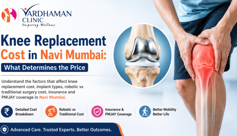
Knee replacement, also called knee arthroplasty, involves replacing a damaged or arthritic knee joint with an artificial implant made of medical-grade metal and plastic. It is one of the most successful orthopedic procedures performed today, with a high long-term success rate. But because it involves surgical expertise, imported implants, hospital infrastructure, and post-operative care, the final bill depends on several moving parts.
Broadly, here is what patients across Nerul, Panvel, Kharghar, Vashi, and CBD Belapur can expect:
- Basic package (traditional technique, standard implant): ₹1,75,000 to ₹2,50,000
- Mid-range package (better implant brand, semi-private room): ₹2,50,000 to ₹3,50,000
- Robotic or navigation-assisted surgery with premium implants: ₹3,50,000 to ₹6,00,000+
These figures are per knee. If both knees need replacement in the same sitting (bilateral knee replacement), the cost naturally increases, though many hospitals offer a discounted combined package.
Key Factors That Determine Knee Replacement Surgery Price
Understanding why prices differ so much helps you compare quotes intelligently rather than simply picking the cheapest option. Here are the main factors that affect knee replacement surgery price in Navi Mumbai.
1. Type of Implant Used
The knee implant cost in India is one of the biggest variables. Implants range from standard domestic brands to premium imported ones from companies like Johnson & Johnson, Zimmer Biomet, Stryker, and Smith & Nephew. Imported implants generally last longer, offer better range of motion, and come with stronger warranties, but they also cost more. A single implant set can range from ₹40,000 for a basic domestic option to over ₹1,50,000 for a high-end imported one.
2. Robotic vs Traditional Technique
This is where many patients get confused, so let’s compare robotic vs traditional knee replacement cost directly.
Traditional knee replacement relies on the surgeon’s manual skill and instrumentation to cut and align the bone. It has been performed successfully for decades and remains a reliable, cost-effective option.
Robotic knee replacement uses computer-assisted, image-guided technology to plan the surgery down to the millimeter before the first incision is made. This improves implant alignment accuracy, can reduce soft tissue damage, and often leads to a smoother recovery. The trade-off is cost. The robotic knee replacement cost in India typically runs ₹80,000 to ₹1,50,000 higher than the traditional approach because of the technology and equipment involved.
Neither option is universally “better” for every patient. Your surgeon should assess your knee anatomy, bone quality, and lifestyle before recommending one over the other, rather than pushing the more expensive option by default.
3. Hospital Category and Room Type
A best orthopedic hospital in Nerul or Panvel with NABH accreditation, modern operation theatres, and a dedicated joint replacement unit will naturally charge more than a smaller nursing home. Room category also matters a lot. General ward, semi-private, private, and deluxe rooms each carry a different daily rate, and since a knee replacement typically involves a 3 to 5 day hospital stay, this adds up.
4. Surgeon’s Experience and Fees
An experienced orthopedic surgeon in Navi Mumbai. who has performed hundreds of successful knee replacements will usually charge a professional fee that reflects that expertise. This is not a place to cut corners. A skilled surgeon reduces the risk of complications, revision surgery, and long recovery times, which ultimately saves money and pain in the long run.
5. Pre-Surgery Tests and Anesthesia
Before surgery, doctors run blood tests, ECG, X-rays, and sometimes an MRI to assess joint damage and confirm you are fit for anesthesia. These diagnostics usually cost between ₹5,000 and ₹15,000 depending on how many tests are needed. Anesthesia and OT charges are typically bundled into the surgical package but are worth asking about specifically.
6. Post-Surgery Physiotherapy and Follow-Up
Recovery does not end when you leave the hospital. The knee replacement recovery cost includes physiotherapy sessions, follow-up consultations, medications, and sometimes home care support. Some hospitals include a set number of physiotherapy sessions in the package, while others charge separately. Always clarify this upfront.
7. City and Locality
Location within Navi Mumbai also plays a role. Knee replacement cost in Panvel can differ slightly from rates in Nerul or Vashi simply because of variations in real estate and operating costs between hospitals in these areas. It is worth comparing two or three centers in your preferred locality before finalizing.
What Is Typically Included in a Knee Replacement Package Cost
One of the most common complaints patients have after surgery is unexpected charges. A transparent knee replacement package cost should ideally include:
- Surgeon and anesthetist fees
- Operation theatre and equipment charges
- The knee implant itself
- Hospital room stay for the standard duration (usually 3 to 5 days)
- Nursing care during admission
- Standard medications during the hospital stay
- Routine post-operative dressing and check-ups
What is often excluded and billed separately:
- Pre-admission diagnostic tests
- Blood transfusion, if required
- ICU stay beyond the standard package (only needed in rare cases)
- Extended physiotherapy after discharge
- Any additional imaging if complications arise
Before you commit to a hospital, ask for a written, itemized estimate. A good clinic will never hesitate to give you this in writing, and if a surgeon’s team is reluctant to break down the numbers, treat that as a red flag.
Knee Replacement Insurance Coverage and Government Schemes
Cost should never be the reason someone continues to suffer from debilitating knee pain, and thankfully there are several ways to manage the financial side.
Health Insurance
Most standard health insurance policies in India, including those from private insurers and corporate group policies, cover knee replacement insurance coverage as part of inpatient hospitalization benefits, provided the policy has completed its waiting period for pre-existing conditions (usually 2 to 4 years for osteoarthritis-related claims). Cashless treatment is available at most network hospitals in Navi Mumbai, which means you may not need to pay upfront at all. It is worth calling your insurer directly to confirm your specific coverage limit and any sub-limits on implants, since some policies cap the amount they will pay for the implant itself.
PMJAY Knee Replacement Coverage
For families covered under Ayushman Bharat, PMJAY knee replacement coverage offers cashless treatment up to ₹5 lakh per family per year at empaneled government and private hospitals. Knee replacement is included in the list of over 1,900 procedures covered under the scheme, and the package is designed to cover the surgery, implant, hospitalization, medicines, and a period of follow-up care with no out-of-pocket cost at empaneled centers. If you or a family member holds an Ayushman card, it is worth checking which hospitals in Nerul, Panvel, or Navi Mumbai are empaneled under PMJAY before choosing where to get treated.
EMI and No-Cost Financing
Many hospitals and clinics now partner with financing companies to offer EMI options, allowing patients to spread the knee replacement surgery price over several months without a heavy upfront burden. If insurance coverage is limited, this can be a practical middle path.
Affordable Knee Replacement in Navi Mumbai: How to Choose Wisely
Searching for affordable knee replacement in Navi Mumbai does not mean choosing the lowest quote blindly. Here is a simple checklist to compare options the smart way:
- Ask for a complete written estimate, not a verbal range.
- Check the surgeon’s experience with your specific implant type and technique, robotic or traditional.
- Confirm what is included in the package versus what will be billed extra.
- Ask about the implant brand and its warranty period.
- Understand the expected hospital stay and whether physiotherapy is bundled in.
- Check hospital accreditation and infection control track record.
- Ask about revision surgery rates, since a lower price sometimes reflects lower-quality implants that wear out sooner.
A slightly higher upfront cost with a trusted surgeon and a genuine, high-quality implant is almost always a better long-term investment than the cheapest available option.
Knee Surgery Cost Comparison: Robotic vs Traditional at a Glance
| Factor | Traditional Knee Replacement | Robotic Knee Replacement |
| Average cost per knee | ₹1,75,000 to ₹2,75,000 | ₹3,00,000 to ₹6,00,000 |
| Precision | Surgeon-dependent, manual alignment | Computer-guided, higher accuracy |
| Recovery time | 4 to 6 weeks for basic mobility | Often slightly faster due to less soft tissue disruption |
| Best suited for | Straightforward cases, cost-conscious patients | Complex deformities, patients wanting maximum precision |
| Implant longevity | 15 to 20 years with good technique | Comparable, with potentially better alignment outcomes |
This knee surgery cost comparison should be discussed directly with your surgeon, since the right choice depends on your knee’s condition, not just your budget.
Why Transparency Matters When Choosing Your Surgeon
The single biggest source of patient anxiety around knee replacement is not the surgery itself. It is the uncertainty around cost. Dr. Abhay Chhallani, a trusted orthopedic surgeon in Navi Mumbai based in Nerul, is known for offering clear, itemized cost estimates before surgery, using imported implants from established manufacturers like Johnson & Johnson and Zimmer, and walking patients through robotic and traditional options honestly, based on what is medically appropriate rather than what is more profitable. For patients in Nerul, Panvel, and the wider Navi Mumbai region who want a best knee replacement surgeon in Nerul with a reputation for straightforward communication, this kind of transparency makes a real difference during an already stressful decision.
If you are exploring your options, the best next step is a detailed consultation where your X-rays, medical history, and lifestyle needs are reviewed, followed by a written cost breakdown tailored to your case rather than a generic online estimate.
Frequently Asked Questions
What is the average knee replacement cost in Navi Mumbai?
Most patients pay between ₹1,75,000 and ₹4,50,000 per knee, depending on implant choice, technique, and hospital category.
Is robotic knee replacement worth the extra cost?
For patients with complex deformities or those who want the highest possible alignment precision, yes. For straightforward cases, traditional knee replacement remains a reliable and more affordable option with excellent outcomes.
Does insurance cover knee replacement surgery in Navi Mumbai?
Most comprehensive health insurance policies cover it after the waiting period for pre-existing conditions is completed. Always confirm implant sub-limits with your insurer beforehand.
Is knee replacement free under PMJAY?
Eligible Ayushman Bharat cardholders can get knee replacement surgery cashless at empaneled hospitals, with no out-of-pocket cost, up to the scheme’s annual family limit of ₹5 lakh.
How long does recovery take after knee replacement?
Most patients can walk with support within a day or two and return to routine activities within 4 to 6 weeks, with full recovery over 3 to 6 months.