Hip pain is something most people push through. A dull ache in the groin, a stiffness that takes time to loosen up in the morning, a slight limp that the family notices before you do. People blame it on age, on the weather, on “overexertion.” And so the months pass – and the damage quietly gets worse.
If you are reading this, you or someone close to you is probably dealing with hip joint pain that has gone on too long. This blog will walk you through everything – the early warning signs, what causes hip joint damage, what treatments are available, and when hip replacement surgery in Mumbai becomes the right decision.
Understanding the Hip Joint
The hip is a ball-and-socket joint – the ball being the top of the thigh bone (femur) and the socket being the cup-shaped hollow in the pelvis (acetabulum). A layer of smooth cartilage covers both surfaces, allowing the joint to move with almost no friction under normal conditions.
When this cartilage wears down – due to arthritis, injury, infection, or loss of blood supply – the bones start grinding against each other. That grinding is what causes the deep, aching pain that no amount of rest seems to fully relieve.
What Causes Hip Joint Damage?
Osteoarthritis
The most common cause of hip joint damage in India. Cartilage gradually breaks down over time, and the joint space narrows. It affects both younger and older patients – not just the elderly. Hip arthritis treatment depends on how advanced the damage is.
Rheumatoid Arthritis
An autoimmune condition where the body’s immune system attacks the joint lining, causing persistent inflammation, pain, and eventual joint destruction.
Avascular Necrosis (AVN)
When blood supply to the femoral head is disrupted – due to steroid use, alcohol, trauma, or unknown reasons – the bone tissue begins to die. AVN is increasingly seen in younger patients in their 30s and 40s. Early stages can sometimes be managed with bone-preserving surgery, but advanced AVN requires hip joint replacement surgery.
Hip Fractures
Falls, road accidents, or weakened bones from osteoporosis can lead to hip fractures. In elderly patients especially, a displaced hip fracture almost always requires surgical treatment – and often a hip replacement.
Hip Dysplasia
A structural problem where the hip socket is too shallow to fully cover the femoral head. This puts uneven stress on the cartilage and leads to early arthritis, often in patients who have no idea the condition existed since childhood.
Post-Infection Arthritis (Septic Arthritis)
A severe joint infection can destroy cartilage rapidly. Even after the infection is treated, the joint damage left behind may eventually require hip replacement treatment.
Early Warning Signs You Should Not Ignore
Most patients who finally walk into our clinic have been dealing with symptoms for one, two, sometimes five years. The pattern is almost always the same – slow progression that gets dismissed until it cannot be ignored anymore.
Here are the signs that your hip joint needs medical attention:
Pain in the Groin, Thigh, or Outer Hip
Hip joint pain is most commonly felt in the groin. It can also radiate to the front of the thigh or the outer side of the hip. Many patients mistake it for a muscle pull or lower back problem. If the pain keeps coming back in the same location – especially after walking or climbing stairs – get an X-ray done.
Morning Stiffness That Takes More Than 30 Minutes to Loosen Up
Waking up with a stiff hip that needs time before you can walk normally is a textbook sign of hip arthritis. As the condition worsens, the stiffness takes longer to ease and starts returning after sitting for even short periods.
Pain at Rest or at Night
When hip pain starts disturbing your sleep or is present even when you are lying down and not putting any weight on the joint, it means the inflammation inside the joint has reached a significant level. This is one of the clearest indicators that surgery may be coming.
Difficulty With Simple Daily Tasks
Putting on shoes and socks, crossing your legs, getting up from a low sofa, stepping in and out of a car, climbing stairs – these are the movements that a damaged hip makes progressively harder. When two or three of these activities become difficult or painful on a daily basis, it is time to consult a hip pain doctor in Mumbai without further delay.
A Limp You Did Not Consciously Develop
The body automatically shifts weight away from a painful joint to protect it. This shows up as a limp – often noticed by family members before the patient themselves. A consistent limp means the hip is structurally compromised.
Leg Length Difference
Advanced hip joint damage can cause the affected leg to appear shorter than the other. Patients notice this when their posture changes, one shoe heel wears out faster, or their gait feels uneven.
Medications Providing Less and Less Relief
If you started with occasional painkillers and have now progressed to daily anti-inflammatory medication – and it is still not enough – your condition has advanced well beyond what medicines alone can manage. This is the body telling you that the underlying structural problem needs to be addressed.
Non-Surgical Hip Pain Treatment Options
Surgery is never the first recommendation. Every patient deserves a proper trial of conservative management. Here are the non-surgical options for hip pain treatment:
Physiotherapy
A structured physiotherapy program strengthens the muscles around the hip, improves joint stability, and reduces the load on damaged cartilage. It is most effective in early to moderate stages of arthritis and helps delay the need for surgery significantly.
Anti-Inflammatory Medications (NSAIDs)
Medications like diclofenac, etoricoxib, or naproxen help reduce joint inflammation and pain. These are useful for managing symptoms but do not stop or reverse joint damage.
Corticosteroid Injections
A steroid injection directly into the hip joint can reduce inflammation significantly and provide relief for weeks to several months. It is a useful bridge treatment – especially for patients who are not yet candidates for surgery or need temporary relief before a planned procedure.
PRP (Platelet-Rich Plasma) Therapy
PRP involves injecting a concentration of the patient’s own growth factors into the joint to promote healing and reduce inflammation. Results vary, and it is most suitable for early to moderate cartilage damage rather than end-stage arthritis.
Hyaluronic Acid Injections
These injections improve the lubrication inside the joint and can reduce pain in mild to moderate arthritis cases. They are not effective once cartilage loss is severe.
Weight Management
For every kilogram of excess body weight, the hip joint carries roughly three to four times that load during walking. Even a modest reduction in weight can meaningfully reduce hip joint pain and slow down the progression of arthritis.
Activity Modification and Walking Aids
Avoiding high-impact activities, using a walking stick on the opposite side of the painful hip, and adjusting daily routines can reduce pain levels significantly in early to moderate stages.
When all these options have been tried genuinely – for weeks or months – and the pain continues to worsen and limit daily life, hip replacement surgery becomes the appropriate and necessary step.
When Is Hip Replacement Surgery Actually Needed?
This is the question every patient asks – and the honest answer is that it depends on the individual, not a fixed number or age.
Hip replacement surgery is recommended when:
Conservative treatments have failed to provide lasting, meaningful relief
Hip pain is present constantly – including at night and at rest
X-rays show severe joint space narrowing or bone-on-bone contact
Daily activities like walking, dressing, and basic movement are significantly impaired
Quality of life, mental health, and sleep are being affected by chronic pain
The patient is medically fit to undergo surgery
Age is not the deciding factor. Patients as young as 35–40 with severe AVN or hip dysplasia undergo successful hip replacement surgery in Mumbai every year. At the same time, patients in their 70s and 80s recover well and regain meaningful independence after surgery.
The goal is not to rush into surgery – it is also not to delay it so long that the muscles waste away, the bone quality deteriorates, and recovery becomes harder.
Types of Hip Replacement Surgery
Total Hip Replacement (THR)
The most commonly performed procedure. Both the damaged femoral head and the worn acetabular socket are removed and replaced with implants – typically a metal stem, a ceramic or metal ball, and a plastic or ceramic socket lining. Total hip replacement surgery delivers outstanding pain relief and long-term results for the vast majority of patients.
Partial Hip Replacement (Hemiarthroplasty)
Only the femoral head is replaced. This is most commonly used after a hip fracture in elderly patients where the socket is still in good condition.
Hip Resurfacing
A bone-conserving alternative where the femoral head is reshaped and capped with a metal covering rather than fully removed. Best suited for younger, male patients with good bone quality and no signs of AVN or osteoporosis.
Minimally Invasive Hip Replacement
Performed through smaller incisions using specialized instruments. This approach reduces blood loss, lowers infection risk, causes less muscle disruption, and allows for faster recovery. Not every patient is a suitable candidate, but when appropriate, it offers clear advantages over traditional open surgery.
Revision Hip Replacement
A second surgery performed when a previous hip replacement implant has worn out, loosened, or developed complications. Revision surgery is more complex and requires a surgeon with specific experience in complex hip reconstruction.
Hip Replacement Surgery Recovery – What to Realistically Expect
Recovery is one of the biggest concerns patients have before surgery. Here is a straightforward timeline based on typical outcomes:
Day 1 to 2 – Standing and First Steps Within 24 hours of surgery, most patients are assisted to stand and take a few steps with support. Early movement is intentional – it reduces the risk of blood clots and starts the recovery process immediately.
Week 1 to 2 – Home With a Walker Most patients are discharged within 3–5 days. You will walk with a walker and manage basic activities at home. Pain is managed well with prescribed medications.
Week 3 to 6 – Increasing Independence Walking distance improves. Many patients transition from a walker to a cane. Physiotherapy continues with progressive exercises to rebuild strength and range of motion.
Month 2 to 3 – Return to Normal Daily Life Most patients are walking without support, sleeping comfortably, and handling all daily activities independently. Driving can usually resume around the 6-week mark as advised by your surgeon.
Month 6 to 12 – Full Recovery Strength, balance, and confidence in the new joint continue to improve. Patients return to activities like swimming, cycling, and light recreational sports.
Hip replacement recovery time is faster and more comfortable than most patients expect – particularly when the physiotherapy program is followed consistently.
Implants Used in Hip Replacement – What You Should Know
Not all hip implants are the same. The choice of implant depends on the patient’s age, weight, activity level, bone quality, and the surgeon’s assessment.
Bearing Surfaces:
Metal on Polyethylene – Most commonly used. Durable and proven over decades.
Ceramic on Ceramic – Very low wear rate, preferred for younger, active patients.
Ceramic on Polyethylene – A good balance of durability and reduced wear.
Fixation Methods:
Cemented – Implant fixed with bone cement. Often preferred for elderly patients with softer bone.
Uncemented (Cementless) – Implant press-fitted into the bone, which then grows into it. Preferred for younger, active patients with good bone quality.
Hybrid – Combination of cemented and uncemented components.
Modern hip replacement implants are designed to last 20–25 years or more. The quality of the implant, combined with the skill of the surgeon and patient compliance in recovery, are the three main factors that determine how long it lasts.
Hip Replacement Surgery in Mumbai – What Patients From Across Maharashtra Need to Know
Mumbai is one of India’s leading centres for joint replacement surgery, with access to advanced implants, modern operation theatres, and experienced orthopedic surgeons. Patients travel from Navi Mumbai, Thane, Pune, Nashik, and other parts of Maharashtra to access this care.
When choosing where to get hip replacement surgery in Mumbai, look for:
A surgeon who performs a high volume of hip replacement procedures annually
A hospital with a dedicated joint replacement unit and post-operative physiotherapy
Transparent communication about implant options, risks, and costs
A care team that takes time to explain your condition before recommending surgery
About Dr. Abhay Chhallani – Orthopedic Surgeon in Mumbai
Dr. Abhay Chhallani is a trusted orthopedic surgeon in Mumbai with extensive experience in hip joint replacement surgery and complex orthopedic conditions. Patients from Mumbai, Navi Mumbai, Thane, Bandra, Andheri, and other parts of Maharashtra consult him for hip pain treatment, hip arthritis management, and joint replacement surgery.
His areas of expertise include:
Total and partial hip replacement surgery
Minimally invasive hip replacement
Avascular necrosis (AVN) treatment and hip preservation
Revision hip replacement surgery
Hip fracture management
Complex cases involving hip dysplasia and post-infection arthritis
As hip replacement doctor in Navi Mumbai and Mumbai, Dr. Chhallani takes a structured approach – exhausting conservative options first, explaining every stage of the decision-making process clearly, and recommending surgery only when it is clinically justified and the patient is fully informed.
Every patient who walks in with hip pain gets a proper examination, imaging review, and a clear explanation of where they stand – and what their options are.
Frequently Asked Questions
Q: I am only 45 years old. Is hip replacement surgery suitable for me?
Yes. Age alone does not determine whether surgery is appropriate. If your hip joint is severely damaged – due to AVN, hip dysplasia, or arthritis – and conservative treatment has failed, hip replacement surgery is the right solution regardless of age. Modern implants are built for active, younger patients.
Q: How long will the implant last?
Current data shows that over 90% of hip replacements last 20 years or more. The right implant choice for your age, weight, and activity level – made by your surgeon – plays a big role in long-term durability.
Q: Is the surgery painful?
The surgery is performed under anaesthesia. Post-operative pain is managed effectively with medications. Most patients are surprised at how manageable the pain is compared to what they expected. Within a few weeks, the post-surgical discomfort reduces substantially.
Q: What activities can I do after hip replacement?
Walking, swimming, cycling, golf, and light recreational activity are all generally permitted. High-impact sports like running or jumping are typically avoided to protect the implant. Your surgeon will give you specific guidance based on your implant type and recovery.
Q: What is the cost of hip replacement surgery in Mumbai?
The cost varies based on the type of implant selected, the hospital, and the surgical approach. Dr. Chhallani’s team provides a full, transparent cost breakdown during your consultation – including implant costs, hospital charges, and post-operative care.
Q: I have been told to wait because I am “too young” for surgery. Is that right?
This is a common – and sometimes outdated – concern. Delaying surgery in a patient with severe joint damage can lead to muscle weakness, bone loss, and a more difficult recovery when surgery finally happens. The right time to operate is when the evidence clearly supports it, regardless of age.
Take the First Step
Living with hip pain every single day wears a person down – physically and mentally. The good news is that hip replacement surgery has transformed the lives of millions of patients, and recovery is faster and more comfortable than it used to be.
If you are in Mumbai, Navi Mumbai, Thane, or anywhere in Maharashtra and you are dealing with hip joint pain that has stopped responding to treatment – do not wait for it to get worse.
Consult Dr. Abhay Chhallani – Hip Pain Doctor in Mumbai and Orthopedic Surgeon for Hip Replacement
📞 Book your consultation today and get a clear, honest assessment of your hip health and your options.
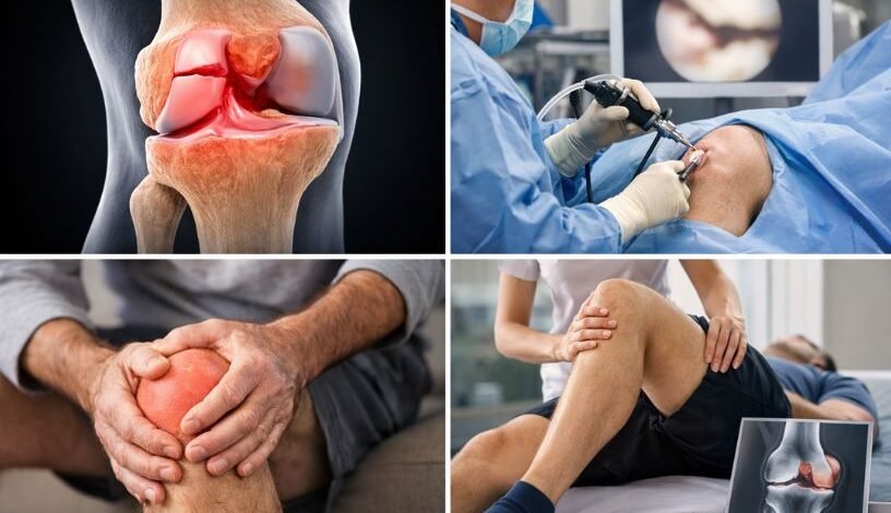
You wake up in the morning. Before your feet even touch the floor, it starts – that deep, grinding ache in your knee. Stairs feel like a test of willpower. A simple walk to the kitchen becomes something you dread. Over time, you stop doing the things that matter most: morning walks, playing with your grandchildren, standing long enough to cook a full meal, or even sitting through a movie without shifting uncomfortably.
If this is your life right now, you are not alone.
Chronic knee pain caused by arthritis, joint degeneration, or old injuries affects millions of people across India – and across Mumbai in particular. For decades, the only lasting solution was traditional total knee replacement, a major surgery with a long recovery that many patients feared.
That has changed.
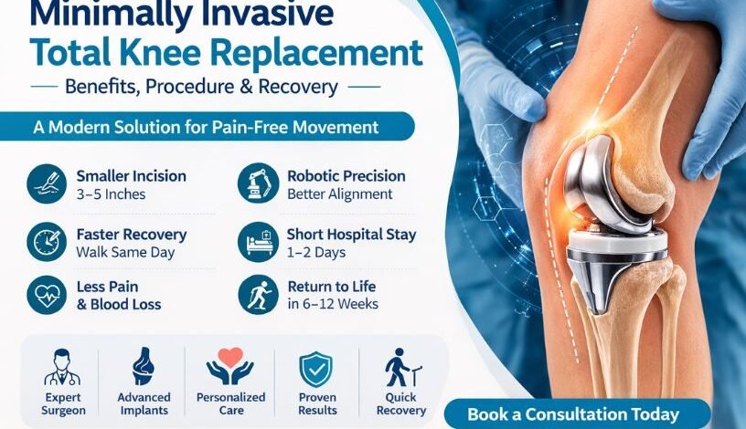
Minimally Invasive Total Knee Replacement (MI-TKR) has emerged as one of the most significant advances in modern orthopedic surgery. It offers the same permanent relief from knee pain as traditional surgery – but with a smaller incision, less damage to surrounding tissue, less post-operative pain, and a faster road back to your normal life.
At our practice, Dr. Abhay Chhallani performs this advanced procedure for patients from across Mumbai, Navi Mumbai, and Maharashtra. As a leading specialist in Robotic Knee Replacement in Navi Mumbai, Dr. Chhallani helps patients get back to walking, moving, and living – often faster than they ever expected.
This comprehensive guide will walk you through everything: what the surgery involves, how it is performed, who is a good candidate, what the different types of implants are, how to prepare before surgery, what recovery looks like week by week, the risks to know about, and how to choose the right surgeon.
What Is Minimally Invasive Total Knee Replacement?
Minimally Invasive Total Knee Replacement – also written as MI-TKR or minimally invasive total knee arthroplasty (MI-TKA) – is an advanced form of the standard total knee replacement procedure.
The fundamental goal is identical to traditional surgery: the damaged surfaces of the knee joint are removed and replaced with precision-engineered metal and high-density plastic implants. This eliminates the bone-on-bone grinding that causes chronic knee arthritis pain and restores smooth, stable joint movement.
What is different is the surgical approach.
In traditional knee replacement surgery, the surgeon makes a large incision of 8 to 12 inches down the front of the knee and fully cuts through the quadriceps tendon – the powerful tendon that connects the thigh muscles to the kneecap. The kneecap is then flipped 180 degrees and the shinbone (tibia) is often dislocated to fully expose the joint.
In minimally invasive knee replacement, the incision is just 3 to 5 inches. Rather than cutting through the quadriceps tendon, the surgeon gently moves the muscles and soft tissue aside. The kneecap is shifted rather than flipped. The tibia is usually left in place.
This approach is known as “quadriceps-sparing” – and it is the key reason why recovery is faster and post-operative pain is lower.
The implants used are identical. Only the technique changes.
Understanding the Knee Joint: Why It Breaks Down
To understand why knee replacement becomes necessary, it helps to understand how the knee works and why it can fail over time.
The knee is the largest and one of the most complex joints in the human body. It connects three bones – the femur (thigh bone), the tibia (shin bone), and the patella (kneecap). The ends of these bones are covered with a smooth, white tissue called articular cartilage, which allows them to glide against each other without friction.
Between the femur and tibia sit two C-shaped pieces of cartilage called the menisci, which act as shock absorbers and help distribute weight evenly across the joint.
When articular cartilage breaks down – due to age, wear and tear, injury, or inflammatory conditions – the bones begin rubbing directly against each other. This causes:
Constant or intermittent pain, especially with movement
Swelling and stiffness in the joint
A grinding, clicking, or locking sensation
Progressive loss of mobility and flexibility
Visible deformity such as the leg bowing inward or outward
Once cartilage is lost, it does not grow back on its own. This is why knee arthritis is progressive – it gets worse over time without treatment, not better.
Types of Knee Conditions That Lead to Replacement Surgery
Not all knee pain leads to replacement surgery, but several specific conditions are its most common causes:
Osteoarthritis (OA)
The most common cause of knee replacement surgery in India. Osteoarthritis is a degenerative joint disease where cartilage gradually wears away over time. It is more common with age, and women are more frequently affected than men due to differences in joint structure and hormonal factors. OA causes progressive pain, stiffness, swelling, and eventually bone-on-bone contact that makes even resting painful.
Rheumatoid Arthritis (RA)
An autoimmune condition in which the body’s immune system attacks the lining of the joint (synovium), causing inflammation, pain, and joint destruction. Rheumatoid arthritis can affect people of any age and often affects multiple joints at once.
Post-Traumatic Arthritis
Develops after a significant knee injury – such as a fracture of the knee bones, a torn ACL (anterior cruciate ligament), or a meniscal tear. Even injuries that appeared to heal well can lead to arthritis years or decades later as the cartilage degrades due to the mechanical disruption caused by the original trauma.
Avascular Necrosis (AVN)
A condition where the blood supply to the bone is cut off, causing bone tissue to die. In the knee, this typically affects the femoral condyle. Without adequate blood supply, the bone weakens and collapses, destroying the joint surface. AVN can be caused by long-term steroid use, excessive alcohol consumption, or prior trauma.
Severe Bone Deformity
Some patients develop significant bowing of the legs – either inward (varus deformity) or outward (valgus deformity) – due to uneven cartilage loss and bone wear. This deformity places abnormal stress on the knee and accelerates joint damage.
Who Needs Total Knee Replacement Surgery?
Total knee replacement surgery is not the first line of treatment – it is recommended only when conservative, non-surgical approaches are no longer providing meaningful relief.
You may be a candidate for total knee replacement surgery if:
You have been diagnosed with severe knee osteoarthritis, rheumatoid arthritis, or post-traumatic arthritis
You experience persistent knee pain that limits your everyday activities – walking, climbing stairs, rising from a chair, or sleeping
Your knee is stiff for more than 30 minutes after waking up or after prolonged sitting
X-rays confirm significant joint space narrowing, bone spurs, or bone-on-bone contact
Your knee has noticeable deformity – bowing in or out
Non-surgical treatments such as pain medication, physiotherapy, steroid injections, PRP therapy, and bracing have failed to provide adequate relief
Your quality of life is seriously compromised by knee pain
You are aged between 55 and 80 (though younger and older patients are also treated when clinically indicated)
The decision to proceed with surgery is never made lightly. It involves a thorough clinical evaluation, detailed imaging, a review of your overall health, and an open conversation between you and your orthopedic surgeon about your goals and expectations.
Non-Surgical Knee Arthritis Treatment Options
Before considering surgery, a well-structured non-surgical treatment plan is always the first step. Understanding these options helps patients know when they have genuinely exhausted conservative management.
Pain and Anti-Inflammatory Medications NSAIDs (non-steroidal anti-inflammatory drugs) such as ibuprofen or diclofenac reduce inflammation and manage pain. These are effective for mild to moderate arthritis but become less reliable as the condition progresses. Long-term NSAID use also carries risks for the stomach, kidneys, and cardiovascular system.
Physiotherapy and Exercise Therapy A structured physiotherapy program strengthens the muscles around the knee – particularly the quadriceps, hamstrings, and calf muscles – which reduces stress on the joint. Low-impact activities like swimming, cycling, and water aerobics are particularly beneficial for knee arthritis patients.
Weight Management Excess body weight dramatically increases the load on the knee joint. Studies show that each kilogram of body weight reduction reduces approximately 4 kilograms of force on the knee. Even modest weight loss can meaningfully reduce pain and slow the progression of arthritis.
Corticosteroid Injections Injections of corticosteroid medication directly into the knee joint can provide significant but temporary pain relief – typically lasting weeks to a few months. Most surgeons limit these to 3 to 4 injections per year to prevent further joint damage from the steroid itself.
Hyaluronic Acid (Viscosupplementation) Injections Hyaluronic acid is a naturally occurring substance in joint fluid that acts as a lubricant and shock absorber. Injectable hyaluronic acid supplements this fluid, potentially reducing pain and improving mobility. Results vary between patients.
Platelet-Rich Plasma (PRP) Therapy PRP is derived from the patient’s own blood. After centrifugation, the platelet-rich fraction is injected into the knee to stimulate tissue healing and reduce inflammation. PRP is increasingly used for early to moderate arthritis and can delay the need for surgery in appropriate patients.
Bracing and Orthotics Knee braces designed for arthritis patients can offload pressure from the damaged compartment of the knee, reducing pain and improving walking comfort. Custom shoe insoles (orthotics) can also correct biomechanical imbalances that worsen knee pain.
When these options no longer provide adequate relief, and when knee pain is significantly impacting your daily function and quality of life, total knee replacement surgery becomes the most effective long-term solution.
Minimally Invasive vs. Traditional Knee Replacement: A Detailed Comparison
Understanding the differences between the two approaches helps patients make informed decisions.
Feature
Traditional TKR
Minimally Invasive TKR
Incision Length
8–12 inches
3–5 inches
Quadriceps Tendon
Cut through
Preserved / gently moved
Kneecap (Patella)
Flipped 180 degrees
Gently shifted to the side
Tibia Dislocation
Often required
Usually not required
Tissue Disruption
Significant
Minimal
Blood Loss
More
Less
Hospital Stay
3–5 days
1–2 days (or same-day)
Early Recovery
Slower
Faster in first 4–6 weeks
Long-Term Outcomes
Excellent
Equally excellent
Implants Used
Proven standard
Same proven standard
Surgical Difficulty
Standard
More technically demanding
Suitable For
Most patients
Carefully selected patients
Important note: The long-term outcomes and implant durability of minimally invasive knee replacement are equivalent to traditional surgery. The advantages are primarily in the early recovery period – less pain, faster return of mobility, and shorter hospital stay.
Types of Minimally Invasive Knee Replacement Surgical Approaches
Orthopedic surgeons use several distinct techniques within the category of minimally invasive knee replacement. Each has slightly different incision placement and muscle handling:
1. Quadriceps-Sparing (QS) Approach
The most conservative minimally invasive technique. The incision avoids the quadriceps tendon entirely. The muscles are retracted – not cut – to access the joint. This approach offers the greatest quadriceps preservation but also provides the most limited surgical field, requiring exceptional skill and specialized instruments.
2. Mini-Midvastus Approach
A small incision is made in the vastus medialis oblique (VMO) muscle – one of the quadriceps muscles on the inner thigh. This gives slightly better access than the quadriceps-sparing technique while still avoiding the main quadriceps tendon. It is the most commonly used minimally invasive approach.
3. Mini-Subvastus Approach
The incision is placed below the vastus medialis muscle rather than through it, going underneath the muscle to reach the joint. This approach fully spares the quadriceps mechanism and can result in excellent early recovery of quad strength, but it is technically more challenging in patients with limited surgical access.
4. Jiffy Knee Technique
A newer, highly specialized approach where only the skin and joint capsule are incised. Specialized instruments gently lift and move the muscles aside without any muscle cutting at all. This technique is performed by very few highly specialized surgeons and is at the frontier of minimally invasive knee surgery.
Your surgeon will determine which approach is most suitable based on your anatomy, body type, and the specific pattern of your knee arthritis.
Types of Knee Replacement Implants
The choice of implant is a critical decision that your surgeon makes based on your age, bone quality, activity level, and the specific anatomy of your knee. Understanding the main types helps patients have more informed conversations with their doctor.
By Fixation Method
Cemented Fixation The most commonly used method worldwide. A fast-curing acrylic bone cement (polymethylmethacrylate) is used to bond the metal implant components to the prepared bone surfaces. Cemented fixation provides immediate, reliable stability and is the gold standard for older patients, those with osteoporosis, or anyone with weaker bone quality.
Cementless Fixation The implant surfaces are coated with a porous or textured material that encourages the patient’s own bone to grow directly into and around the implant over time. This biological bonding can provide outstanding long-term durability. Cementless implants are generally preferred for younger, more active patients with good bone quality. The bone ingrowth process takes several weeks to months to complete. Recent research and large registry studies show that cementless knee replacement achieves survivorship rates exceeding 98% at 5 years.
Hybrid Fixation A combination approach where the tibial (shin bone) component is cemented and the femoral (thigh bone) component is cementless, or vice versa. Used when bone quality varies between the two surfaces.
By Bearing Design
Fixed-Bearing Implants The polyethylene (plastic) spacer is fixed firmly to the metal tibial tray and does not move independently. These are durable, reliable, and well-suited for less active patients or those with simpler knee mechanics.
Mobile-Bearing Implants (Rotating Platform) The polyethylene spacer can rotate a small amount within the metal tibial tray, mimicking the natural rotational movement of the knee. This design can improve range of motion slightly and reduce wear on the plastic component. Mobile-bearing implants are often preferred for younger, more active patients who require more natural knee kinematics.
By Constraint Level
Cruciate-Retaining (CR) Design The posterior cruciate ligament (PCL) is preserved during surgery. This design aims to maintain a more natural feel in the knee and can improve proprioception (the sense of joint position). Suitable when the PCL is intact and functioning.
Posterior-Stabilized (PS) Design A post-and-cam mechanism built into the implant replaces the function of the PCL, which is removed during surgery. PS designs provide more reliable stability and are preferred when the PCL is damaged, incompetent, or removed.
Your surgeon will choose the implant design that best matches your anatomy, lifestyle, and long-term goals.
Robotic Knee Replacement Surgery: The Future of Precision
One of the most exciting developments in knee replacement surgery is the integration of robotic-assisted technology with the minimally invasive approach.
Robotic knee replacement surgery – also known as robotic-assisted total knee arthroplasty – combines the soft-tissue-sparing benefits of minimally invasive surgery with extraordinary implant placement accuracy that goes beyond what can be consistently achieved with manual instrumentation alone.
Patients seeking Robotic Knee Replacement in Navi Mumbai now have access to this world-class technology close to home, with Dr. Abhay Chhallani offering robotic-assisted procedures using the latest surgical platforms available in the region.
How Robotic Knee Replacement Works
Before surgery, a detailed CT scan of the patient’s knee is used to create a precise three-dimensional model of the joint. Using this model, the surgeon and the robotic system together develop a fully customized surgical plan – specifying exactly where bone cuts will be made, what size implant will be used, and the ideal alignment and positioning of each component.
In the operating room, the robotic arm provides haptic feedback and real-time guidance to the surgeon. The arm can physically limit bone cuts to the pre-planned boundaries, making it virtually impossible to remove more bone than intended. This level of precision is particularly valuable in minimally invasive surgery, where the reduced field of vision makes accuracy more challenging.
Why Accuracy Matters So Much in Knee Replacement
The long-term success of a knee replacement depends heavily on how well the implant is aligned. Even a few degrees of misalignment can accelerate wear on the plastic spacer, reduce range of motion, cause ongoing pain, and ultimately shorten the life of the implant – potentially requiring revision surgery.
Robotic assistance helps ensure:
Optimal implant alignment and positioning
Correct leg axis and balance
Precise soft tissue balancing
Personalized implant sizing based on the patient’s exact anatomy
Computer-Assisted Navigation
For practices without robotic arms, computer-assisted navigation systems provide real-time 3D feedback during surgery using infrared sensors attached to the bones. Systems like OrthoPilot and Knee Track Module allow surgeons to verify alignment and implant position continuously during the procedure, significantly improving accuracy over conventional manual techniques.
Patient-Specific Instrumentation (PSI) Another technology where custom-designed surgical guides are manufactured from pre-operative CT or MRI scans. These guides fit the patient’s unique bone anatomy like a key in a lock, directing the exact location and angle of each bone cut. PSI reduces operating time and enhances reproducibility.
How to Prepare for Minimally Invasive Knee Replacement Surgery
The weeks and months before your surgery are just as important as the surgery itself. Patients who are well-prepared physically and mentally tend to recover faster and experience fewer complications.
4 to 8 Weeks Before Surgery: Pre-Habilitation Exercises
Pre-habilitation – strengthening the muscles around your knee before surgery – is one of the most evidence-backed ways to improve recovery outcomes.
Recommended exercises before knee replacement:
Quad Sets Lie flat on your back with your leg straight. Tighten the thigh muscle (quadriceps) and press the back of your knee into the floor or bed. Hold for 5 seconds, then relax. Repeat 10 times, twice daily.
Straight Leg Raises Lie on your back. Bend your non-operated leg with foot flat. Tighten the thigh of the operated leg and raise it to the height of the opposite knee. Hold for 3 seconds, lower slowly. Repeat 10 times.
Heel Slides Lie on your back. Slowly slide your heel toward your buttocks, bending the knee as far as comfortable. Hold for 5 seconds, then slide back. Repeat 10 times. This improves knee flexion range pre-operatively.
Short Arc Quads Place a rolled towel under your knee. Straighten your leg fully, hold 5 seconds, lower slowly. Repeat 10 times. Excellent for isolated quad strengthening.
Seated Knee Flexion and Extension Sit in a chair and bend and straighten your knee through its comfortable range of motion. Repeat 10–15 times. Especially beneficial before surgery to maintain range.
Standing Hip Abduction Stand holding a stable surface. Lift your leg out to the side, keeping it straight and toes forward. Hold 3 seconds, lower slowly. This strengthens the hip stabilizers, which support the knee.
Low-Impact Cardiovascular Exercise Stationary cycling, swimming, and water walking are excellent options before knee replacement. They build cardiovascular fitness, reduce pain, manage weight, and maintain muscle tone – all without placing excessive stress on the damaged joint.
Nutrition Before Knee Replacement Surgery
What you eat in the weeks before surgery directly affects your healing, immunity, and recovery speed.
High Protein Diet Aim to eat a high-quality protein source 3 to 4 times per day in the month before surgery. Protein is the building block of muscle and tissue repair. Good sources include chicken, fish, eggs, paneer, dal, lentils, and Greek yogurt.
Iron-Rich Foods Spinach, lentils, red meat, and fortified cereals support healthy red blood cell production, which is important both during surgery and healing.
Vitamin C Supports collagen production and wound healing. Found in citrus fruits, guava, amla, bell peppers, and tomatoes.
Calcium and Vitamin D Support bone health and strength. Dairy products, fortified foods, leafy greens, and safe sun exposure support calcium and vitamin D levels.
Fiber Constipation is a common side effect of post-operative pain medications (opioids). A fiber-rich diet before and after surgery – from whole grains, fruits, and vegetables – helps prevent this issue.
Hydration Stay well-hydrated in the days before surgery. Avoid alcohol for at least two weeks pre-operatively, as it can interfere with anesthesia, increase bleeding, and impair healing.
Other Pre-Surgery Preparation Steps
Stop Smoking Smoking significantly impairs healing by reducing blood flow and oxygen delivery to tissues. It also increases the risk of infection and wound complications. Stopping smoking at least 6 to 8 weeks before surgery substantially reduces these risks.
Medication Review Inform your surgeon about every medication and supplement you take – including blood thinners (aspirin, warfarin, clopidogrel), NSAIDs, diabetes medications, herbal supplements, and vitamins. Many of these need to be paused or adjusted before surgery.
Medical Clearance A pre-operative assessment with your physician will evaluate your heart health, lung function, blood sugar control (especially important for diabetic patients), and blood pressure. Any uncontrolled medical condition needs to be optimized before surgery can safely proceed.
Pre-Operative Tests Blood tests, electrocardiogram (ECG), chest X-ray, and knee imaging (X-rays and sometimes MRI) are typically required before surgery.
Home Preparation Arrange your home before surgery to make your recovery smoother:
Remove loose rugs and tripping hazards from floors
Set up a ground-floor sleeping area if stairs are involved
Install grab bars in the bathroom
Arrange a raised toilet seat if possible
Stock up on easy-to-prepare meals for the first few weeks
Arrange for someone to help you at home for the first 1 to 2 weeks
If you have children or pets, arrange appropriate care
How Is Minimally Invasive Knee Replacement Surgery Performed? (Step-by-Step)
Pre-Operative Preparation
On the day of surgery, you will fast for 6 to 8 hours beforehand. An intravenous (IV) line will be placed, and antibiotic medication is given before the incision to prevent infection.
Anesthesia
The procedure is performed under general anesthesia (you are fully unconscious) or spinal anesthesia (you are awake but numb from the waist down). Many patients also receive a nerve block – an injection of local anesthetic around the knee nerves – which significantly reduces post-operative pain for the first 12 to 24 hours and reduces the need for strong opioid pain medications.
Tourniquet Application
A tourniquet is inflated around the upper thigh to temporarily reduce blood flow to the knee during the procedure. This reduces intraoperative blood loss and provides a clearer surgical field.
The Minimally Invasive Incision
A precise incision of 3 to 5 inches is made over the front of the knee. Using specially designed narrow retractors, the surgeon carefully moves the quadriceps muscle and soft tissue aside – rather than cutting through them.
Exposing the Joint
The kneecap (patella) is gently moved to one side rather than flipped over, limiting the disruption to the extensor mechanism of the knee. Specialized instruments allow the surgeon to work within the smaller field of view.
Bone Preparation
The surgeon precisely removes the damaged cartilage and a thin layer of underlying bone from the end of the femur, the top of the tibia, and if required, the undersurface of the patella. Specially designed minimally invasive cutting guides – or robotic/computer assistance – ensure these bone cuts are made at exactly the right angles.
Trial Components
Before cementing the permanent implants, trial components are placed to test the fit, alignment, range of motion, and ligament balance. This is one of the most critical steps – ensuring the knee feels natural and balanced before the final implants are secured.
Implant Fixation
The permanent metal femoral and tibial components are fixed using bone cement, cementless press-fitting, or a combination of both – depending on the patient and the implant selected. A high-quality polyethylene spacer is snapped between the two metal components. If the patella is being resurfaced, a plastic button is cemented to its undersurface.
Irrigation and Closure
The joint is thoroughly washed out. A drain may be placed to collect fluid post-operatively. The layers of tissue are carefully closed in sequence, and the skin is closed with sutures or staples.
Total Procedure Time
The entire surgery typically takes 1.5 to 2.5 hours, depending on complexity.
Key Benefits of Minimally Invasive Knee Replacement Surgery
1. Smaller Incision and Less Visible Scar
At 3 to 5 inches, the incision is less than half the length of traditional surgery. Patients – particularly those conscious about surgical scars – appreciate this significantly.
2. Less Post-Operative Pain
The quadriceps-sparing approach means less tissue is cut and less healing is required of the surrounding muscles. Much of the intense pain after traditional knee replacement comes from the healing of the divided quadriceps tendon – which is largely avoided here. This translates to lower opioid requirements post-surgery, which itself reduces side effects like nausea, constipation, and drowsiness.
3. Faster Early Recovery
Because the quadriceps are preserved, muscle function returns faster. Patients are typically walking with a walker on the same day as surgery and often progress to a cane within 2 to 3 weeks – faster than with traditional surgery.
4. Shorter Hospital Stay
Most patients undergoing minimally invasive knee replacement are discharged after 1 to 2 days. Carefully selected patients can even be discharged the same day as surgery. This reduces hospital costs and the risk of hospital-acquired infections.
5. Less Blood Loss
The smaller incision and reduced tissue handling naturally result in less bleeding during surgery, reducing the likelihood of needing a blood transfusion.
6. Lower Infection Risk
Smaller incisions mean less wound surface area exposed during surgery and less dead tissue to become infected. This is especially meaningful for patients with diabetes, obesity, or conditions that slow healing.
7. Faster Return of Knee Range of Motion
Patients with preserved quadriceps muscles tend to achieve better early range of motion in physiotherapy, which is directly linked to better long-term outcomes.
8. Earlier Return to Daily Life
Patients typically return to light daily activities in 2 to 4 weeks, driving in 4 to 6 weeks, and office work in 3 to 6 weeks.
9. Equivalent Long-Term Outcomes
The long-term durability and success rates of minimally invasive knee replacement are equivalent to traditional knee replacement. Modern knee implants have survivorship rates exceeding 90 to 95% at 10 years – meaning most patients get a decade or more of excellent, pain-free function before any further intervention is needed.
Understanding the Risks and Complications
Any surgical procedure carries risks, and it is important that patients have an honest understanding of what these are. The overall rate of serious complications following total knee replacement – minimally invasive or traditional – is low, typically less than 5 percent when performed by an experienced, high-volume surgeon.
Possible complications include:
Blood Clots (Deep Vein Thrombosis / DVT) One of the most common post-operative concerns. Blood clots can form in the deep veins of the leg after surgery. To prevent this, patients are started on blood-thinning medications and encouraged to mobilize early. Signs include increased leg swelling, redness, and warmth.
Pulmonary Embolism (PE) A blood clot that travels to the lungs. This is a serious complication but is rare when proper DVT prevention measures are taken.
Infection Infection around the knee implant is a rare but serious complication. Surgeons administer antibiotics before, during, and after surgery to minimize this risk. Patients are advised to inform dentists and other doctors about their knee replacement before any invasive procedures in future, as bacteria from elsewhere in the body can travel to the implant.
Implant Loosening or Misalignment Over years or decades, implants can loosen from the bone or shift slightly out of alignment. Modern implant design, robotic assistance, and careful patient selection have reduced this risk significantly.
Nerve and Blood Vessel Injury The nerves and blood vessels around the knee are at small risk of injury during surgery. Experienced surgeons using specialized minimally invasive instruments and modern technology minimize this risk.
Stiffness (Arthrofibrosis) Some patients develop excessive scar tissue inside the knee joint after surgery, leading to stiffness and limited range of motion. Consistent physiotherapy is the best preventive measure.
Implant-Specific Complications
Cementless implants may have a period of slight pain until bone ingrowth is complete
Mobile-bearing implants carry a very small risk of polyethylene dislocation
Anesthesia Risks Like all surgeries requiring anesthesia, there are small risks related to cardiac events, stroke, and reactions to anesthesia agents. Pre-operative clearance and a thorough anesthesia evaluation minimize these risks.
Overall: The risks of knee replacement surgery must always be weighed against the significant quality-of-life benefits for patients who have exhausted all conservative treatment options. The vast majority of patients experience significant, lasting improvement in pain and mobility.
Minimally Invasive Knee Replacement: Who Is – and Is Not – a Candidate?
Good Candidates
Generally healthy patients aside from their knee condition
Moderate to severe knee arthritis without extreme bone loss
No prior major open knee surgery on the same knee
BMI under approximately 35 (excess weight makes the limited access more technically difficult)
No severe knee deformity (moderate deformity can often still be addressed)
Intact or only mildly compromised ligaments
Patients Who May Need Traditional Surgery Instead
Significant obesity (high BMI) that limits surgical access
Severe bone deformity requiring more extensive correction
Previous major open knee surgery with significant scar tissue
Very severe bone loss requiring structural bone grafting
Complex revision (redo) knee replacement cases
Medical conditions that significantly increase surgical risk
The decision between minimally invasive and traditional surgery is made individually for every patient. What matters most is not the size of the incision – but that the surgery is performed correctly, with well-positioned implants, by a surgeon with the training and experience to deliver consistent results.
Week-by-Week Recovery Guide After Minimally Invasive Knee Replacement
Day of Surgery (Day 0)
Within hours of waking from anesthesia, a physiotherapist will visit you. Under guidance, you will sit on the edge of the bed, and most patients take their first steps with a walker on the same day. The operated leg is elevated to reduce swelling. Ice packs are applied regularly. IV pain medication transitions to oral medications.
Days 1–3: Early Recovery in Hospital
The focus shifts to controlled walking, going up and down steps (a required skill before discharge), independent transfer in and out of bed, beginning basic knee exercises (quad sets, heel slides, ankle pumps), and wound care.
Ankle pumps – repeatedly flexing and pointing the foot – are done every hour while resting. This activates the calf muscles and helps prevent blood clots.
Week 1–2: Home Recovery Begins
You will likely use a walker or forearm crutches. The priority is managing swelling (elevation and ice packs), wound hygiene, and performing your prescribed home exercise program 2 to 3 times per day.
Most patients experience their worst swelling during this period – this is completely normal. Swelling can persist for weeks to months as the body heals.
A physiotherapist visits at home or you attend outpatient sessions – this varies based on your recovery plan.
Week 3–6: Building Range of Motion and Strength
By week 3, most patients can walk with a cane or independently for short distances. Physiotherapy intensifies – targeting knee flexion beyond 90 degrees, full knee extension, stair climbing technique, and progressive muscle strengthening.
Driving is generally not recommended during this period, especially if the operated leg is needed for braking.
Most patients can resume light cooking, personal hygiene without assistance, and short outings.
Week 6–12: Growing Independence
The 6-week milestone is significant. Most patients have noticeably reduced pain, better walking endurance, and improved confidence in their knee. Low-impact activities like swimming and stationary cycling are typically introduced at this stage.
Office-based workers often return to work in this period. Driving is usually permitted after clearance from your surgeon, typically around 6 weeks.
3 to 6 Months: Return to Normal Life
By 3 months, the majority of patients feel dramatically better than before surgery. Pain is minimal or absent during most activities. Physiotherapy continues but decreases in frequency.
Activities like golf, light hiking, dancing, cycling, and recreational walking are generally achievable by this point for most patients.
6 Months to 1 Year: Full Recovery
Complete recovery – including the final return of full strength and stability – takes up to 12 months. Mild swelling at the end of the day can persist for up to a year after surgery. This is normal.
Most patients at 1 year are living the active, pain-free lives they underwent surgery to achieve.
Physiotherapy: The Single Most Important Factor in Recovery
Physiotherapy is not optional after knee replacement. It is essential.
A dedicated and consistent physiotherapy program:
Restores knee range of motion (bending and straightening)
Rebuilds quadriceps strength and muscle control
Retrains the nervous system for safe, coordinated movement
Prevents stiffness and scar tissue formation
Reduces the risk of long-term complications
Helps patients return to activities sooner
The standard physiotherapy program after knee replacement progresses in phases:
Phase 1 (0–6 weeks): Pain and swelling management, basic range of motion, safe transfers, walking re-education with walking aids
Phase 3 (3–6 months): Functional training specific to the patient’s goals – whether that means returning to gardening, dancing, playing with children, or low-impact sports
Phase 4 (6–12 months): Activity-specific conditioning and full return to active lifestyle
Patients who attend all physiotherapy sessions and consistently perform their home exercise program have measurably better outcomes than those who do not.
Life After Knee Replacement: Activities and Expectations
Understanding what you can realistically expect from life after knee replacement helps patients set appropriate goals.
Activities generally suitable after knee replacement:
Walking (including long-distance walks and hiking on even terrain)
Swimming and water aerobics
Cycling (stationary and road cycling)
Golf
Dancing
Light recreational sports
Travel, including long-haul flights (with appropriate DVT precautions)
Yoga and stretching
Activities to approach with caution:
Jogging and running (high-impact, may accelerate implant wear)
Racquet sports involving sudden direction changes (badminton, squash)
High-impact aerobics classes
Contact sports
Activities generally to avoid after knee replacement:
High-impact jumping activities
Heavy weightlifting with deep knee bends
Skiing (downhill)
Contact sports (football, wrestling)
How long do knee replacement implants last? Modern knee replacement implants have a proven track record of lasting 15 to 20+ years with proper care. Studies consistently show survivorship rates exceeding 90% at 10 years and around 80 to 85% at 20 years. Younger patients and those with higher activity levels may wear out implants faster.
Will I need a revision surgery? Most patients never need revision (redo) surgery. However, implants can eventually loosen, wear out, or develop complications requiring revision. Your orthopedic surgeon will monitor your knee with periodic follow-up appointments.
Conclusion: A Pain-Free Future Is Possible
Knee pain does not have to define the rest of your life. With minimally invasive total knee replacement, backed by modern implant technology and robotic precision, thousands of patients each year in India discover that returning to an active, comfortable, and fulfilling life is not just possible – it is achievable faster than they ever expected.
The right surgery, performed by the right surgeon, with the right preparation and the right rehabilitation, can genuinely transform your life.
If you are living with knee arthritis, chronic knee joint pain, or knee deformity in Mumbai or Navi Mumbai – take the first step today.
Book a consultation with Dr. Abhay Chhallani – trusted specialist in Robotic Knee Replacement in Navi Mumbai – and find out whether minimally invasive knee replacement is the right solution for you.
Why Choose Dr. Abhay Chhallani for Knee Replacement Surgery in Mumbai?
Choosing the right surgeon is the single most important decision in your knee replacement journey. The minimally invasive technique is technically demanding – it requires specialized instruments, years of dedicated training, a high case volume, and the expertise to handle unexpected complexity without compromising the result.
Dr. Abhay Chhallani is recognized as one of the best orthopedic surgeons in Mumbai and Navi Mumbai, with extensive experience in minimally invasive and robotic knee replacement surgery, complex joint reconstruction, and revision knee arthroplasty. For patients specifically looking for Robotic Knee Replacement in Navi Mumbai, Dr. Chhallani’s practice offers one of the most advanced and accessible options in the region.
Patients from across Mumbai, Thane, Navi Mumbai, Pune, and beyond choose Dr. Chhallani because of:
Deep specialization in minimally invasive total and partial knee arthroplasty
Robotic and computer-navigated surgery for exceptional implant precision
Individualized treatment planning – every surgical plan is customized to the patient’s unique anatomy and goals
Comprehensive pre-operative preparation programs including physiotherapy, nutritional guidance, and medical optimization
Dedicated rehabilitation team ensuring structured, guided recovery from day one
Transparent, patient-centered consultations where all options are fully explained with realistic expectations
Strong outcomes and consistent patient satisfaction from years of specialized knee replacement practice
Convenient Mumbai location with access to world-class surgical facilities
Whether you are in the early stages of exploring your options or ready to schedule surgery, Dr. Chhallani’s team is here to guide you through every step of the process – from first consultation to full recovery.
A meniscal tear is one of the most common causes of persistent knee pain across all age groups. From young athletes and gym enthusiasts to middle-aged office workers and elderly individuals, this injury can significantly affect mobility and long-term joint health. With increasing participation in sports, rising obesity rates, and more sedentary lifestyles weakening joint structures, cases of meniscal tear are steadily increasing.
The biggest concern patients face is whether they truly need surgery or if conservative knee injury treatment is enough. Many people mistake temporary pain relief for actual healing. Pain may reduce with medications, rest, or physiotherapy, but the internal tear may still remain unstable.
Choosing the wrong treatment—or delaying necessary surgery—can accelerate cartilage damage and lead to early arthritis. That’s why understanding when surgery is needed is essential for protecting your knee for the long term.
Understanding the Meniscus: Why It Is So Important
Before deciding about surgery, it’s important to understand the structure involved.
The meniscus is a C-shaped cartilage cushion between the thigh bone (femur) and shin bone (tibia). Each knee has two menisci:
Medial meniscus (inner side)
Lateral meniscus (outer side)
Their functions include:
Shock absorption
Joint stability
Load distribution
Lubrication support
Protection of knee cartilage
When the meniscus tears, the knee loses its ability to distribute weight evenly. This increases stress on cartilage and can lead to long-term degeneration if untreated.
Meniscal Tear vs General Knee Pain: Why Proper Diagnosis Matters
Knee pain is one of the most common orthopedic complaints. However, not all knee pain is caused by a meniscus injury.
Why Knee Pain Is Often Misdiagnosed
Many patients assume swelling and discomfort mean arthritis or simple strain. However, knee pain causes are diverse and may include:
Ligament tears (ACL, MCL injuries)
Patellar tracking disorders
Tendinitis
Early osteoarthritis
Cartilage defects
Meniscal tears
Symptoms like stiffness, clicking, swelling, or instability overlap between conditions. Without proper evaluation, treatment may target the wrong problem.
Conditions That Mimic Meniscal Tears
Several issues resemble meniscal tears:
Patellofemoral pain syndrome
Baker’s cyst
Synovial inflammation
Loose cartilage fragments
Minor ligament sprains
Accurate diagnosis requires:
Detailed clinical examination
Special orthopedic tests
MRI imaging when needed
A specialist evaluation ensures the correct treatment pathway.
Types of Meniscal Tears and Their Severity
Not all tears are the same. The type and pattern of tear strongly influence whether surgery is required.
Common meniscus tear types include:
Horizontal tear – Often degenerative
Vertical tear – May be repairable
Radial tear – Disrupts shock absorption
Bucket-handle tear – Causes locking
Flap tear – Creates unstable fragments
Complex tear – Multiple tear patterns combined
Tears in the outer “red zone” (good blood supply) may heal with conservative treatment. Tears in the inner “white zone” (poor blood supply) rarely heal naturally and often need surgery.
Patient Profile Matters: Who Is More Likely to Need Surgery?
The decision for surgery depends heavily on the individual.
Age and Activity Level
Young Athletes: Sports involving twisting (football, basketball, cricket, tennis) often cause traumatic tears. These tears are usually larger and unstable, increasing the likelihood of surgery.
Middle-Aged Adults: Degenerative tears develop gradually due to wear and tear. Some can be managed without surgery if symptoms are mild.
Elderly Patients: If arthritis is already present, surgery decisions become more complex and individualized.
Manual laborers cannot function with mechanical instability.
Activity demands significantly influence meniscus tear surgery candidates.
Timeline-Based Decision Making for Meniscal Tears
Time-based monitoring is crucial in deciding surgery.
First 0–2 Weeks: Acute Phase
Initial management includes:
Rest and ice therapy
Anti-inflammatory medication
Temporary activity restriction
Knee brace support
Minor tears may settle during this phase.
Weeks 3–6: Rehabilitation Phase
Physiotherapy focuses on:
Quadriceps strengthening
Hamstring conditioning
Balance exercises
Range-of-motion restoration
If symptoms significantly improve, surgery may not be required.
Beyond 6–8 Weeks: Evaluation Window
If pain, swelling, or mechanical symptoms persist beyond 6–8 weeks despite proper therapy, surgical consultation becomes important.
The typical meniscus tear recovery time without surgery ranges from 4–8 weeks for stable tears. Persistent symptoms suggest structural instability.
Red Flag Symptoms That Strongly Indicate Surgery
Certain symptoms strongly favor surgical treatment:
1. True Mechanical Locking
If the knee gets stuck and cannot move temporarily, it indicates internal obstruction.
2. Inability to Fully Straighten the Knee
A displaced fragment may block extension.
3. Recurrent Swelling
Swelling after minimal activity signals ongoing irritation.
4. Sharp Twisting Pain
Pain during rotation movements suggests unstable tear fragments.
Symptoms like knee locking are classic indicators of a severe meniscus tear requiring surgical attention.
Tear Characteristics That Require Surgical Intervention
Surgery becomes more likely when:
The tear is displaced
The tear causes joint obstruction
The tear is in the avascular zone
The tear pattern is complex
The knee shows repeated instability
These tears cannot heal properly with rest alone.
Impact of Delaying Surgery: What Can Go Wrong?
An untreated meniscus tear can lead to:
Progressive Cartilage Damage
Without shock absorption, cartilage wears out faster.
Early Knee Arthritis
Joint degeneration accelerates significantly.
Muscle Weakness and Instability
Chronic pain reduces activity, leading to muscle loss.
Reduced Repair Success
Delay may convert a repairable tear into one needing removal.
Early decision-making improves outcomes.
Surgery vs Non-Surgical Care: Detailed Comparison
Factor
Non-Surgical Care
Surgical Care
Pain Relief
Gradual
Faster for mechanical symptoms
Knee Stability
Limited in unstable tears
Restores stability
Recovery Time
4–8 weeks (mild tears)
2–6 weeks (partial removal)
Arthritis Prevention
Limited
Better in selected cases
Long-Term Protection
Depends on tear type
Better for unstable tears
Understanding available meniscus treatment options helps patients choose wisely.
Types of Meniscal Surgeries and When Each Is Chosen
Most procedures are done using minimally invasive arthroscopic surgery.
Meniscus Repair
Tear is stitched
Preserves cartilage
Ideal for younger patients
Longer rehabilitation
Partial Meniscectomy
Damaged portion removed
Faster recovery
Used when repair not possible
Surgeons consider:
Tear location
Tissue quality
Patient age
Activity demands
The goal is to preserve as much meniscus as possible.
Recovery Expectations Based on Surgery Type
Weight Bearing
Repair: Limited for 4–6 weeks
Partial removal: Immediate walking possible
Return to Work
Office job: 1–3 weeks
Physical work: 6–12 weeks
Return to Sports
Repair: 4–6 months
Partial removal: 6–8 weeks
Strict physiotherapy is essential for optimal meniscus surgery recovery.
Cost, Hospital Stay, and Practical Considerations
Hospital Stay
Most surgeries are daycare procedures.
Meniscus Surgery Cost
The meniscus surgery cost depends on:
City and hospital category
Surgeon experience
Type of procedure
In India, costs generally range between ₹70,000 – ₹1,80,000.
Insurance
Most insurance policies cover arthroscopic procedures if medically necessary. Pre-authorization may be required.
Can Surgery Be Avoided Permanently?
In selected cases, yes.
Early Diagnosis
Early intervention prevents worsening.
Muscle Strengthening
Strong thigh muscles reduce knee stress.
Weight Management
Excess body weight increases knee load.
Activity Modification
Avoid deep squats and sudden twisting.
Understanding how to prevent meniscus surgery involves proactive care and strengthening.
Frequently Asked Patient Questions
What happens if I delay surgery?
Delay may increase cartilage damage and arthritis risk.
Will surgery completely cure the tear?
It resolves mechanical symptoms but requires rehabilitation for best results.
Can I live with a meniscal tear?
Yes, if stable and minimally symptomatic.
How do surgeons decide during arthroscopy?
They directly assess tear stability and blood supply before choosing meniscus repair or removal.
Conclusion: Making an Informed Decision About Meniscal Surgery
There is no universal answer to whether surgery is needed for a meniscal tear treatment plan. The decision depends on:
Patient age
Activity level
Tear type
Symptom severity
Response to conservative treatment
Early evaluation by an orthopedic specialist ensures accurate diagnosis and personalized treatment. Acting at the right time protects your knee from long-term degeneration and helps maintain an active, pain-free life.
If you are experiencing persistent knee pain, swelling, or locking, do not ignore the symptoms. A timely expert opinion can prevent serious complications and safeguard your joint health for years to come.
Hip pain is one of the most common musculoskeletal complaints seen in adults and elderly individuals. However, even younger people are increasingly experiencing hip discomfort due to sedentary lifestyles and prolonged sitting. Living with hip pain can slowly affect independence, confidence, and mobility.
In the early stage, hip pain may appear only during certain activities like walking long distances or climbing stairs. Over time, it can progress into chronic hip pain, affecting everyday movements such as bending, standing, or even getting out of bed.
Early consultation and proper hip pain treatment can prevent further joint deterioration. In severe degenerative cases, advanced surgical options such as hip replacement in Navi Mumbai may be recommended to restore joint function and eliminate persistent pain.
Understanding Hip Pain
What Is Hip Pain and Where It Occurs
The hip joint is a ball-and-socket joint that supports body weight and allows smooth movement. Hip joint pain usually occurs in the groin area but may also radiate to the thigh, buttocks, or lower back.
Understanding pain location helps identify the cause:
Deep groin pain → Often joint-related
Outer hip pain → Usually bursitis or tendon inflammation
Buttock pain → May indicate nerve-related issues
Pain radiating down the leg → Possible sciatica
Acute vs Chronic Hip Pain
Acute hip pain develops suddenly due to injury, fall, or muscle strain.
Chronic hip pain lasts more than 6 weeks and is often linked to arthritis or long-term wear and tear.
Daily habits like crossing legs while sitting, poor sleeping posture, weak core muscles, and obesity significantly contribute to the causes of hip pain.
Common Causes of Hip Pain in Daily Life
Understanding the major hip pain reasons helps in choosing appropriate treatment.
Age-Related Causes
Osteoarthritis of the hip
Most common cause in older adults
Cartilage breakdown leads to bone friction
Causes stiffness and grinding sensation
Wear and tear of the hip joint
Reduced joint lubrication
Decreased bone density
In advanced arthritis, when conservative treatments fail, doctors may suggest hip replacement in Navi Mumbai to improve mobility and relieve long-term pain.
Lifestyle & Activity-Related Causes
Modern lifestyle plays a major role in the causes of hip pain, including:
Sitting for 8–10 hours daily
Working on non-ergonomic chairs
Lack of regular stretching
Sudden intense workouts without warm-up
High-impact sports like running on hard surfaces
Weak hip muscles and tight hip flexors increase joint pressure, leading to chronic discomfort.
Medical & Injury-Related Causes
Hip bursitis (inflammation of fluid-filled sacs)
Hip labral tear (damage to cartilage ring)
Muscle strain or ligament sprain
Sciatica-related hip pain
Rheumatoid arthritis
Avascular necrosis
These conditions may cause sharp, burning, or deep aching pain, depending on severity.
Daily Challenges Faced While Living With Hip Pain
Hip Pain While Walking
Walking becomes uncomfortable due to:
Reduced stride length
Limping
Increased pain after 10–15 minutes
Difficulty walking on uneven surfaces
Over time, abnormal walking patterns can affect knees and lower back.
Hip Pain While Sitting
Prolonged sitting may lead to:
Hip tightness
Numbness in thighs
Pain when standing after sitting
Reduced flexibility
Office workers often experience worsening hip pain in daily life due to continuous sitting.
Hip Pain While Climbing Stairs
Climbing stairs puts extra load on the hip joint, leading to:
Sharp pain
Weakness
Knee compensation
Loss of balance
Hip Pain at Night and While Sleeping
Night pain is common in chronic hip pain patients. It may cause:
Frequent waking
Inability to lie on affected side
Sleep deprivation
Daytime fatigue
Poor sleep can worsen pain sensitivity.
Impact on Work and Mental Health
Long-term hip pain can cause:
Reduced concentration
Anxiety about movement
Social withdrawal
Fear of surgery
Decreased overall quality of life
Chronic pain often affects emotional well-being along with physical health.
Warning Signs That Hip Pain Should Not Be Ignored
Knowing when to see a doctor for hip pain is important. Seek medical advice if:
Pain persists beyond 2–3 weeks
You feel joint locking or catching
There is visible swelling
You cannot bear weight
Pain radiates below the knee
Early medical intervention can reduce the need for major surgical procedures later.
Diagnosis of Hip Pain
Accurate hip pain diagnosis ensures proper treatment.
An orthopedic doctor for hip pain may recommend:
Detailed medical history evaluation
Physical movement tests
X-ray to detect arthritis
MRI for ligament, tendon, or labral injuries
CT scan in complex cases
Blood tests for inflammatory arthritis
Timely diagnosis helps prevent worsening joint damage and reduces the chances of needing hip replacement in Navi Mumbai.
Hip Pain Treatment Options
Non-Surgical Hip Pain Treatment
Most early-stage patients improve with hip pain treatment without surgery, including:
Anti-inflammatory medications
Muscle relaxants
Structured physiotherapy
Strength training exercises
Hydrotherapy
Physiotherapy strengthens hip abductors and stabilizing muscles, reducing joint stress.
Lifestyle-Based Treatment for Daily Relief
Effective hip pain daily care includes:
Avoid sitting for more than 30–40 minutes continuously
Using lumbar and hip support cushions
Sleeping on a medium-firm mattress
Losing excess body weight
Using shock-absorbing footwear
These changes provide sustainable hip pain relief.
Advanced Medical Treatments
If conservative care fails:
Corticosteroid injection therapy for hip pain
PRP injections to promote healing
Hyaluronic acid injections
Radiofrequency ablation in selected cases
These treatments can delay or prevent surgery.
Surgical Treatment
When Surgery Becomes Necessary
Surgery is considered when:
Severe osteoarthritis is confirmed
Pain limits daily activities
Non-surgical treatment fails
Joint deformity progresses
Hip Arthroscopy
Minimally invasive procedure to repair labral tears and remove damaged tissue.
Hip Replacement Surgery
In severe arthritis or joint destruction, hip replacement surgery is the most effective solution. Patients seeking advanced orthopedic care often opt for hip replacement in Navi Mumbai, where modern surgical techniques, minimally invasive approaches, and structured rehabilitation programs improve recovery outcomes.
Daily Life Tips for Managing Hip Pain
Proper hip pain management requires consistency.
Safe Exercises
Glute bridges
Hip rotations
Gentle hamstring stretches
Water-based exercises
Activities to Avoid
Deep squats
High-impact jumping
Sudden twisting movements
Sitting cross-legged for long durations
Prevention of Hip Pain in Daily Life
To prevent hip pain, focus on:
Regular strengthening exercises
Maintaining ideal body weight
Practicing yoga for flexibility
Taking standing breaks at work
Correct lifting techniques
Proper hip pain prevention reduces long-term joint damage risk.
Recovery and Long-Term Care
Expected Recovery Time
Mild conditions → 4–6 weeks
Moderate injuries → 8–12 weeks
After hip replacement in Navi Mumbai → 3–6 months full recovery with rehabilitation
Role of Physiotherapy
Physiotherapy is crucial for hip pain recovery, improving mobility, balance, and muscle strength.
Preventing Recurrence
Continue strengthening exercises
Maintain ergonomic posture
Regular follow-ups with orthopedic specialist
Conclusion: Living Better Despite Hip Pain
Effective hip pain relief begins with early detection and proper hip pain treatment. Ignoring symptoms can lead to long-term joint damage and mobility loss.
With timely diagnosis, lifestyle improvements, physiotherapy, and when required, advanced solutions like hip replacement in Navi Mumbai, patients can regain comfort, mobility, and confidence.