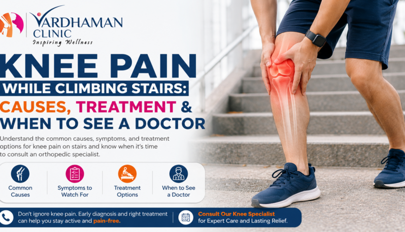
That familiar twinge on the third or fourth step. The sharp pinch when you push off the landing. The dull ache that follows you all the way to the top floor. If knee pain while climbing stairs has become a regular part of your day, you are not alone, and more importantly, you should not ignore it.
Knee pain on stairs is one of the most commonly reported orthopedic complaints, yet one of the most frequently brushed aside. People assume it is age, or weather, or just the price of being busy. But pain in knee when climbing stairs is often the first clear signal that something is happening inside the joint that deserves attention. Conditions like osteoarthritis knee disease, knee cartilage damage, patellofemoral pain syndrome, and meniscus tear symptoms may feel manageable on flat ground but become noticeably worse the moment you hit a staircase.
Why do my knees hurt when I climb stairs? The answer lies in physics. Climbing stairs forces the knee to bend more deeply and absorb significantly more load than walking on flat ground. The force across the kneecap during stair climbing can be three to four times your body weight. Any underlying damage or inflammation in the joint gets amplified under that load, which is exactly why do my knees hurt when I climb stairs even when the rest of the day feels manageable.
In this blog, Dr. Abhay Chhallani explains the most common causes of knee joint pain on stairs, what your symptoms may be telling you, available knee pain treatment options, and the signs that mean it is time to see an orthopedic doctor for knee pain without further delay.
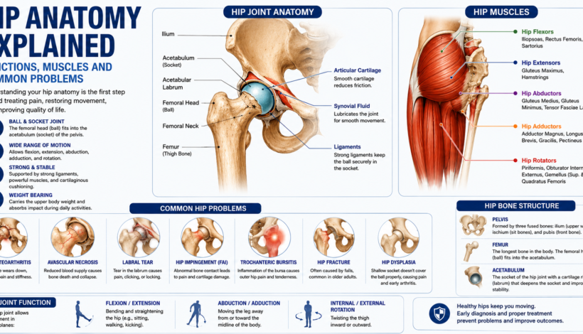
Why Stairs Are Hard on the Knee Joint
Before getting into causes, it helps to understand what the knee is actually doing when you climb stairs.
With each step up, the quadriceps muscle contracts powerfully to lift your entire body weight against gravity. This pulls on the patella, pressing it firmly into the groove at the base of the femur. The cartilage behind the kneecap, the menisci between the thigh and shin bones, and the surrounding ligaments all absorb and redistribute that force simultaneously.
On level ground, that system works smoothly even when mildly irritated. On stairs, the increased bend angle and the added load expose whatever weakness or damage exists in the joint. This is why knee pain while walking upstairs often appears before pain shows up during walking, and why stairs are such a reliable early indicator of joint problems.
Common Causes of Knee Pain While Climbing Stairs
1. Patellofemoral Pain Syndrome
Patellofemoral pain syndrome is the single most common cause of knee pain on stairs, particularly in younger and middle-aged patients.
It occurs when the kneecap does not track properly in its groove on the thigh bone. Instead of gliding smoothly, it shifts slightly to one side, creating uneven pressure and irritation on the cartilage beneath it. The result is knee joint pain centred around or behind the kneecap, which gets significantly worse during stair climbing, squatting, or prolonged sitting.
Patients often describe a dull ache at the front of the knee that builds with activity. Some notice a grinding sensation or a soft click during movement. The pain tends to ease with rest but returns quickly on the next flight of stairs.
Patellofemoral pain syndrome responds well to physiotherapy, especially exercises that strengthen the quadriceps and hip abductors to improve kneecap tracking. Most patients see meaningful improvement within six to eight weeks of consistent rehabilitation.
2. Osteoarthritis of the Knee
Osteoarthritis knee disease is the most common cause of knee pain while climbing stairs in patients over 45.
In osteoarthritis, the cartilage that lines the joint surfaces gradually wears away. As it thins, the cushioning between bones reduces and the joint becomes more sensitive to load. Stairs, which create several times the force of flat walking, trigger knee arthritis symptoms that may be barely noticeable on level ground.
Knee arthritis symptoms on stairs typically include pain and stiffness after the first few steps, a grinding or grating sensation in the joint, and significant difficulty descending stairs, which is often worse than climbing. Knee swelling that appears after activity is also common.
The progression of osteoarthritis knee disease is slow, but it does not reverse. Early management with physiotherapy, weight control, anti-inflammatory medication, and injections can slow the decline and reduce symptoms significantly. When conservative treatment stops working, surgical evaluation becomes appropriate.
3. Knee Cartilage Damage (Chondromalacia Patella)
Knee cartilage damage specifically on the underside of the kneecap, a condition called chondromalacia patella, is closely related to patellofemoral syndrome but involves actual softening and breakdown of the cartilage itself.
The cartilage behind the kneecap is not equipped to handle abnormal or repetitive stress well. When it softens or develops surface irregularities, stair climbing becomes painful because of the direct compression between the kneecap and the femur at each step.
Patients with knee cartilage damage often report a roughness or grating sensation under the kneecap, sensitivity when pressing on the front of the knee, and knee stiffness after sitting with the knee bent for long periods. Getting up from a chair and then immediately climbing stairs is particularly uncomfortable.
Knee cartilage damage is diagnosed with an MRI scan, which shows the extent and location of the softening. Treatment depends on severity and ranges from physiotherapy and activity modification to cartilage repair procedures in younger patients with isolated damage.
4. Meniscus Tear
Meniscus tear symptoms on stairs often include a sharp, catching pain on one side of the knee, a sense of the knee wanting to give way, and sometimes a distinct clicking or locking sensation mid-step.
Each knee has two menisci, C-shaped discs of cartilage that cushion the joint and distribute load between the thigh bone and the shin bone. When a meniscus tears, whether from a sudden twist during sport or gradually through age-related degeneration, the torn fragment can catch during movement and create localised, intense pain.
Meniscus tear symptoms are often side-specific. Pain on the inner side of the knee points to the medial meniscus; pain on the outer side suggests the lateral meniscus. Stair climbing aggravates both because of the bending and weight-bearing combination required with each step.
Acute tears in younger patients often respond well to surgery, specifically arthroscopic repair. Degenerative tears in older patients are frequently managed without surgery first, using physiotherapy and injections, with good results in many cases.
5. Ligament Injuries
Ligament injuries, particularly to the anterior cruciate ligament or the medial collateral ligament, can make pain in knee when climbing stairs sharp and accompanied by a sense of instability.
Ligaments stabilise the knee during movement. When they are partially or fully torn, the knee has to work harder to maintain stability during load-bearing activities. Stair climbing, which requires coordinated support from all the stabilising structures, puts direct demand on injured ligaments.
Patients with ligament injuries often describe the knee as feeling unreliable, as though it might give way on the step. This is different from the ache of arthritis or the front-of-knee pain of patellofemoral syndrome, and it usually has a more sudden onset following an injury event.
6. Patellar Tendinitis
The patellar tendon runs from the kneecap down to the shin bone. It is central to stair climbing because it is the tendon the quadriceps uses to extend the knee and drive you upward with each step.
When the patellar tendon is irritated or inflamed from overuse, knee pain while climbing stairs is sharp and localised just below the kneecap. The pain is typically worse during and immediately after activity, then eases with rest, only to return on the next bout of stair climbing.
This condition is common in people who have recently increased their physical activity, changed footwear, or spend long periods on their feet. It responds well to physiotherapy focused on tendon loading exercises and activity modification.
7. Bursitis
Small fluid-filled sacs called bursae sit around the knee joint to reduce friction between bones, tendons, and skin. When a bursa becomes inflamed, usually from repetitive pressure or direct impact, the resulting swelling and pain can make knee pain while climbing stairs significant.
Prepatellar bursitis causes swelling directly over the kneecap. Pes anserine bursitis, which affects the inner side of the knee just below the joint, is particularly common in patients with osteoarthritis knee disease and often causes pain on stair descent rather than ascent.
Knee swelling from bursitis has a characteristic boggy, soft feel different from the joint swelling of arthritis. It usually responds well to rest, ice, compression, and anti-inflammatory medication. Persistent cases may require a corticosteroid injection.
Symptoms That Tell You Which Direction to Look
The location and character of your stair pain is a useful diagnostic clue before you even see a doctor.
Front of the knee, around or behind the kneecap: Points to patellofemoral pain syndrome or knee cartilage damage. Very common, often bilateral.
Inner side of the knee: Suggests medial meniscus involvement or medial collateral ligament strain. Often sharp and catching.
Outer side of the knee: Points to lateral meniscus or iliotibial band involvement.
General, deep joint pain: More typical of osteoarthritis knee disease. Often accompanied by knee swelling and knee stiffness in the morning.
Worse going down than going up: Descending stairs creates even more compression across the kneecap and patellofemoral joint. If going down hurts significantly more, cartilage damage or patellofemoral issues are the likely culprits.
Sudden sharp catch mid-step with a giving-way sensation: More consistent with a meniscus tear or ligament instability than arthritis.
Knee Pain Treatment Options
The right knee pain treatment depends entirely on the underlying cause. A correct diagnosis comes first, which is why seeing a knee specialist early matters. That said, here is an overview of the approaches used across conditions.
Physiotherapy and Exercise Rehabilitation
This is the foundation of treatment for nearly every cause of knee pain while climbing stairs. Strengthening the quadriceps, hip abductors, and gluteal muscles improves the mechanics of kneecap tracking, reduces load on the joint during stair climbing, and corrects movement patterns that contribute to pain.
Physiotherapy is not just about the exercises themselves. A trained therapist assesses how you walk, how you climb stairs, and where the weakness or tightness in the kinetic chain is contributing to your knee joint pain. Manual therapy, taping techniques, and gradual load progression are all part of a structured rehabilitation programme.
Most patients with patellofemoral syndrome, mild to moderate osteoarthritis knee disease, and patellar tendinitis see significant improvement with consistent physiotherapy over six to twelve weeks.
Anti-inflammatory Medications
NSAIDs such as ibuprofen or diclofenac reduce both knee swelling and pain during flare-ups. They are useful for short-term relief but not a long-term solution for chronic knee pain caused by structural damage.
Topical anti-inflammatory gels are an effective option for localised pain with fewer systemic side effects, particularly in older patients or those with stomach sensitivity.
Corticosteroid Injections
For persistent knee joint pain that is not responding to physiotherapy and medication, a corticosteroid injection directly into the joint or the affected bursa can provide significant relief, typically lasting two to four months.
This gives the patient a window of reduced pain during which physiotherapy becomes more effective. Injections are not a cure, and repeated injections over time can have downsides, so they are used selectively rather than as a routine first step.
Hyaluronic Acid Injections
Hyaluronic acid, a substance naturally present in joint fluid, can be injected into the knee to improve lubrication and reduce friction in patients with osteoarthritis knee disease. Results are variable but some patients experience meaningful improvement in knee pain while climbing stairs and knee pain while walking upstairs for six months or more.
PRP (Platelet-Rich Plasma) Therapy
PRP therapy involves injecting a concentrated preparation of the patient’s own platelets into the joint to promote healing and reduce inflammation. It is used increasingly for knee cartilage damage and early arthritis. Evidence is growing, though results vary between patients.
Knee Bracing and Orthotics
A patellar stabilising brace with a cutout for the kneecap helps guide proper patellar tracking during stair climbing in patients with patellofemoral syndrome. Hinged braces provide stability for ligament injuries. Foot orthotics address overpronation that can contribute to abnormal knee mechanics.
Surgical Options
Surgery is considered when conservative treatment has failed and symptoms are significantly affecting daily life.
For meniscus tear symptoms that do not resolve, arthroscopic surgery to repair or trim the torn portion is a short, effective procedure with fast recovery. For isolated knee cartilage damage in younger patients, cartilage repair or regeneration procedures are available. For advanced osteoarthritis knee disease, a partial or total knee replacement eliminates the damaged joint surface entirely.
When to See an Orthopedic Doctor for Knee Pain
Many people wait too long. The earlier an orthopedic doctor for knee pain identifies the underlying cause, the more treatment options are available and the better the outcome.
See a knee specialist without delay if:
The pain has lasted more than four to six weeks without clear improvement. Persistent knee pain on stairs is not something to wait out indefinitely.
Swelling is present most of the time, not just after activity. Persistent knee swelling indicates ongoing inflammation inside the joint.
The knee has locked, given way, or buckled on the stairs or otherwise. This suggests structural instability that needs evaluation.
The pain is worsening progressively rather than staying stable. Escalating chronic knee pain means the underlying condition is advancing.
You have stopped using stairs or modified your daily routine to avoid pain. When the joint is dictating your lifestyle choices, it is time to seek an assessment.
There was a specific injury event that preceded the pain, such as a twist, fall, or impact. This raises the possibility of a meniscus tear or ligament injury that benefits from timely diagnosis.
Morning knee stiffness lasts more than 30 to 45 minutes, which is a classic feature of knee arthritis symptoms that deserves evaluation.
A consultation with the best knee doctor involves a clinical examination, assessment of your movement pattern, and usually an X-ray to assess joint space. An MRI is added when soft tissue structures like the meniscus, cartilage, or ligaments need detailed evaluation.
Self-Care Tips While You Wait for Your Appointment
These measures will not fix the underlying problem, but they reduce pain and prevent it from worsening in the short term.
Modify, do not completely avoid. Avoiding stairs entirely weakens the quadriceps further, which makes stair climbing harder over time. Use an elevator during acute flare-ups, but keep doing gentle strengthening exercises.
Ice after activity for 15 to 20 minutes to reduce knee swelling and local inflammation following stair climbing.
Use the handrail and lead with your stronger leg when climbing, your painful leg when descending. This distributes load more favourably.
Maintain a healthy body weight. Each kilogram of body weight adds approximately four kilograms of force across the knee on stairs. Weight reduction has a disproportionate impact on knee joint pain during load-bearing activities.
Wear supportive footwear. Flat, unsupportive shoes worsen knee mechanics. Proper footwear with cushioning and arch support reduces the load transmitted up through the joint.
Knee Treatment in Navi Mumbai
If knee pain while climbing stairs is affecting your daily life and you have been putting off getting it properly evaluated, this is the right time to act.
Dr. Abhay Chhallani offers comprehensive assessment and management for all causes of knee joint pain, from patellofemoral pain syndrome and meniscus tear symptoms to osteoarthritis knee disease and knee cartilage damage. Whether you need physiotherapy guidance, injections, or surgical evaluation, the goal is to identify the exact cause of your pain and give you a clear treatment path.
For knee treatment in Navi Mumbai, contact Dr. Abhay Chhallani’s clinic to book your consultation. Do not let stair pain quietly reshape your life.
Frequently Asked Questions
Is knee pain while climbing stairs always serious?
Not always. Occasional mild discomfort after unusual activity can be normal. But pain that is persistent, worsening, or accompanied by swelling, instability, or significant stiffness should be evaluated by a knee specialist.
Can knee pain on stairs improve without surgery?
Yes, in the majority of cases. Physiotherapy, weight management, medication, and injections resolve symptoms effectively for most conditions. Surgery is considered when conservative treatment over an adequate period has not provided relief.
Why does going downstairs hurt more than going up?
Descending creates greater compression force across the kneecap and patellofemoral joint. If downstairs is significantly more painful than climbing, patellofemoral pain syndrome or knee cartilage damage is often the explanation.
How long does it take to recover from knee pain caused by stairs?
It depends entirely on the cause. Patellofemoral syndrome with physiotherapy often improves in six to eight weeks. Meniscus tears requiring surgery typically have a recovery of four to six weeks. Arthritis management is ongoing rather than curative, focused on reducing symptoms and slowing progression.
When should I see the best knee doctor for stair pain?
If the pain has lasted more than four to six weeks, if the knee is swollen or unstable, if you have changed your daily habits to avoid stairs, or if there was a specific injury, book an appointment with an orthopedic doctor for knee pain for a proper evaluation.